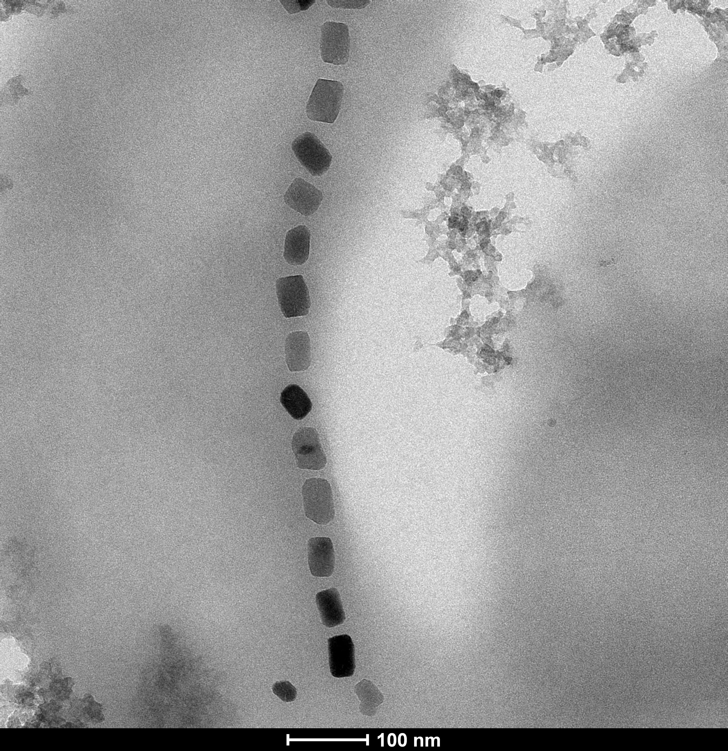
Imagine a tiny vehicle with a nanomagnetic structure, which can be steered through the human body via external magnetic fields. Arriving at its destination, the vehicle may release a drug, or heat up cancer cells without affecting healthy tissue. Scientists of different disciplines are working on this vision. A multidisciplinary research group at Universidad del País Vasco, Leioa, Spain, explores the talents of so-called magnetotactic bacteria, which have the surprising property of forming magnetic iron oxide nanoparticles inside their cells. These particles, with diameters of around 50 nanometers (100 times smaller than blood cells), arrange, within the bacterium, into a chain. The Spanish team is pursuing the idea of using such "magnetic bacteria" as magnetic hyperthermia agents to treat cancer: Steered to the cancer site, the magnetic nanostructures are to be heated by external fields in order to burn the cancer cells.
Now, the researchers have cooperated with a team of physicists led by Sergio Valencia at HZB to explore these magnetic properties in detail. The degree of success for all these applications depends on the magnetic properties of the individual nanomagnets. But since the signals originating from these super tiny magnetic structures are quite weak, it is necessary to average values over thousands of such structures in order to get meaningful data.
Average values are not enough
Until now, only these averaged values could be measured, which puts some constraints in the design of customized nanomagnet applications. But this has now changed. Spanish physicist Lourdes Marcano has developed a new method. "We can now obtain precise information on the magnetic properties of several individual nanomagnets in a simultaneous way," she says.
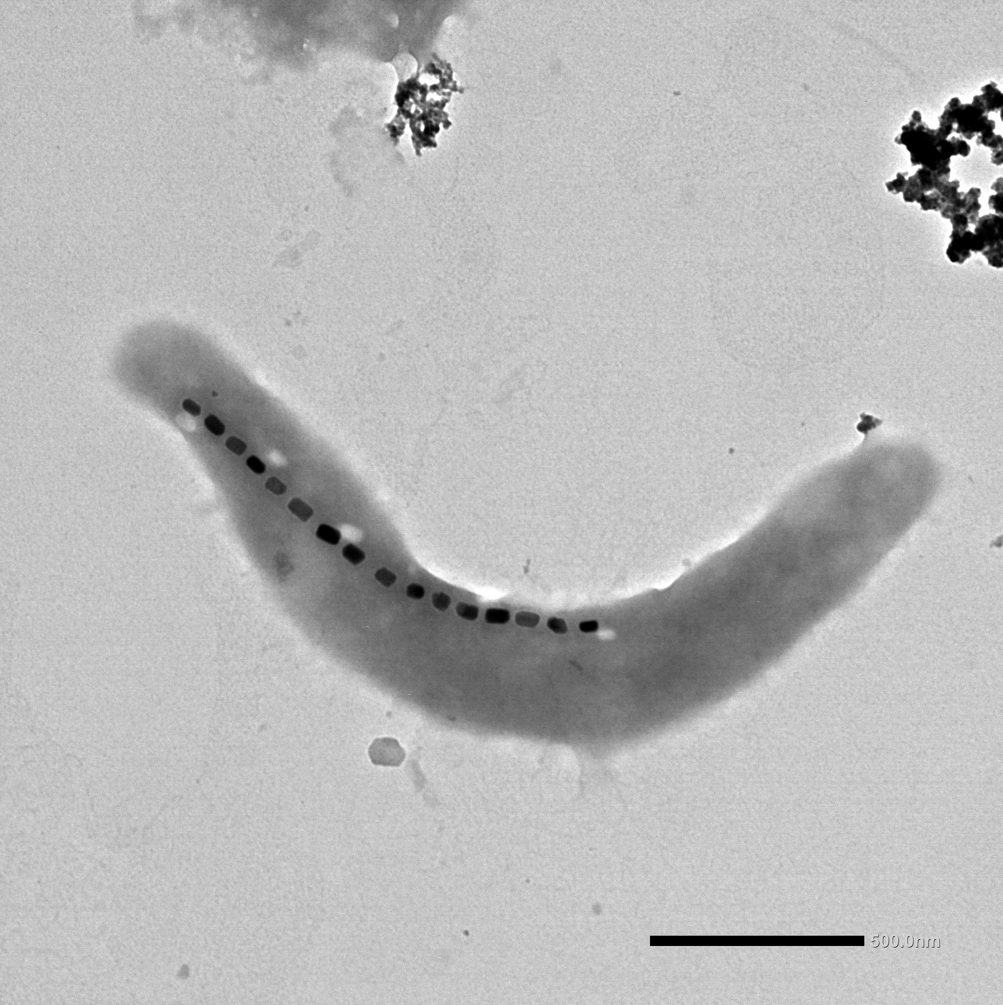
Magnetic anisotropy for every single particle
The method allows measurement of the magnetic properties of individual magnetic nanostructures, even when embedded within biological entities. Magnetic imaging at the scanning transmission X-ray microscope MAXYMUS at BESSY II with the help of theoretical simulations provides information about the so-called magnetic anisotropy of each single nanoparticle within the field of view of the microscope. The method has been proven by determining the magnetic anisotropy of magnetic nanoparticles inside a bacterium. The magnetic anisotropy is an important parameter for controlling and steering magnetic nanoparticles as it describes how a magnetic nanoparticle reacts to external magnetic fields applied at an arbitrary direction.
Future standard lab technique
"Actually, magnetic imaging of magnetic nanoparticles inside a biological cell with enough spatial resolution requires the use of X-ray microscopes. Unfortunately, this is only possible at large scale research facilities, like BESSY II, providing sufficiently intense X-ray radiation. In the future, however, with the development of compact plasma X-ray sources, this method could become a standard laboratory technique," says Sergio Valencia. The research was published in ACS Nano.
Helmholtz Association of German Research Centres
Reference
Magnetic Anisotropy of Individual Nanomagnets Embedded in Biological Systems Determined by Axi-asymmetric X-ray Transmission Microscopy
Lourdes Marcano, Iñaki Orue, David Gandia, Lucía Gandarias, Markus Weigand, Radu Marius Abrudan, Ana García-Prieto, Alfredo García-Arribas, Alicia Muela, M. Luisa Fdez-Gubieda, and Sergio Valencia
https://doi.org/10.1021/acsnano.1c09559
© 2022 American Chemical Society
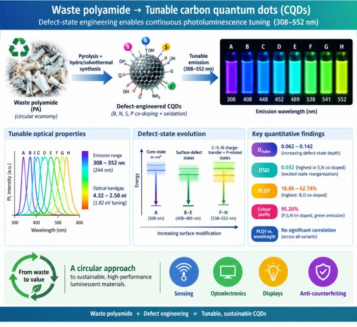
Waste polyamide-derived CQDs show continuous photoluminescence tuning from 308 to 552 nm through sequential defect-state engineering, with optical transition energies decreasing from 4.32 to 2.50 eV. @Christian Ebere Enyoh from Saitama University Carbon quantum dots (CQDs) are fluorescent carbon nanomaterials with potential applications in sensing, optoelectronics, displays, anti-counterfeiting, and environmental technologies. Their optical properties can be adjusted by modifying the carbon...
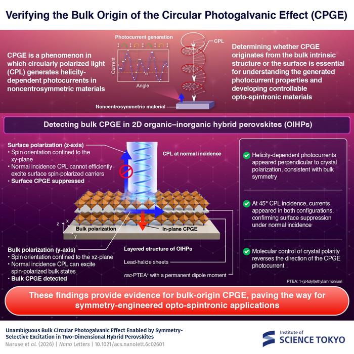
The researchers used circularly polarized light at normal incidence and found that helicity-dependent photocurrents appeared perpendicular to the crystal’s polarization but vanished when measured parallel to it, supporting a bulk origin of CPGE. @Institute of Science Tokyo The circular photogalvanic effect (CPGE), a phenomenon that generates helicity-dependent photocurrents in noncentrosymmetric materials, can originate purely from a crystal's internal structure without contribution from the...
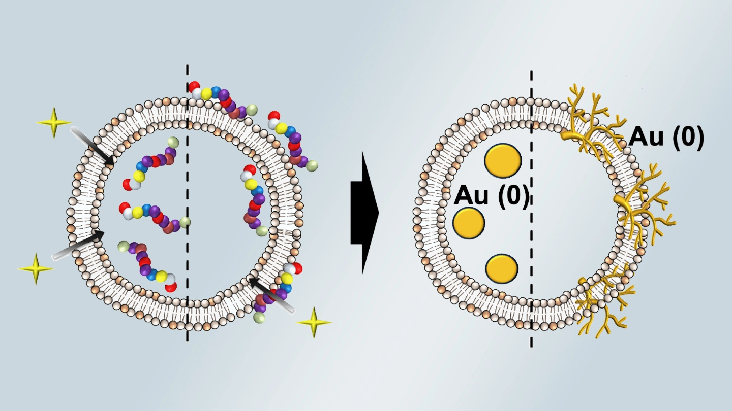
Summary of gold nanoparticle growth programming in liposome based on localization of biomineralization peptide The position of biomineralization peptides within liposomes can influence how gold nanoparticles grow, reports a research team from Institute of Science Tokyo. Peptides localized at the membrane interface promote branched structures, while those confined to the liposome interior favor spherical nanoparticles. The findings offer a new strategy for controlling nanoscale reaction...
