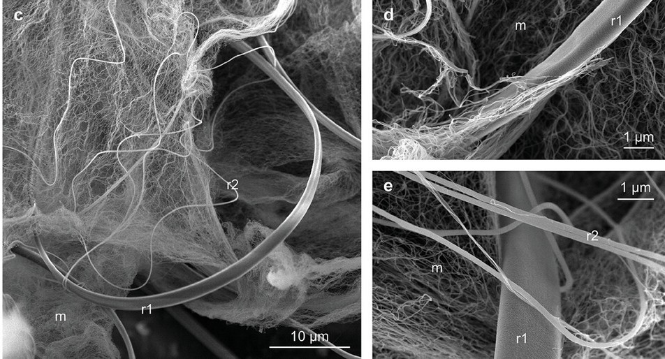
How small is “small?” Spider silk nanofibrils are just a few molecular layers thick, equivalent to approximately one ten-thousandth the diameter of a human hair. They’re invisible to the naked eye and cannot be seen under an ordinary microscope.
Applied science doctoral candidate Jake Silliman ’22 recently measured the strength and stretchability of these miniscule nanofibrils, a feat that his advisor, long term researcher of spider silk VMEC Professor Hannes Schniepp, had previously considered nearly impossible.
“Other people have tried,” Schniepp said. “Most gave up, but Jake didn’t, and he succeeded. If you understand a little bit about what it takes to do what he did, it’s really impressive. It’s actually kind of crazy to think that it’s even possible.”
Innovative research like this highlights the promise of W&M’s new School of Computing, Data Sciences & Physics. Additionally, the research partnership between Silliman and Schniepp serves as an example of the university’s efforts to provide the most personal education of any public university in the United States.
“It’s been one of my greater missions to come up with new materials and new technologies that are inspired by nature,” said Schniepp, who has been studying spider silk for nearly 15 years. “We humans think we’re so great and we can invent things, but if you just take a step outside, you find so many things that are more exciting.”
By weight, spider silk is approximately five times stronger than steel, but Schniepp points out that what makes the material even more impressive is its extensibility. The combination of strength and stretchiness allows it to absorb large amounts of energy. If humans find a way to replicate the structure of spider silk, it could be manufactured for use in practical applications.
“You could make a super bungee cord from it,” said Schniepp. “Or a shield around a structure where you have something incoming at high velocity and you need to absorb a lot of energy. Things like that.”
Spider web strands may seem simple to the casual observer, but closer inspection reveals that they’re complex structures of interwoven components. Schniepp explained that there are approximately 50,000 different species of spiders, and each produces its own unique silk. The silk of the southern house spider was used for this experiment due to the nanofibril mesh found in its structure.
The spinning apparatus of the southern house spider consists of hundreds of nozzles that, with the help of the spider’s back legs, constructs a complicated, three-dimensional strand. The core of the strand is composed of two distinct warps that form helical loops around a central foundation fiber. The tiniest fibers, nanofibrils, are simultaneously spun into a mesh that surrounds those supporting structures.
While webs of some spiders capture prey by way of glue drops spaced regularly along strands, cribellate strands like those spun by the southern house spider ensnare prey in the mesh by way of entanglement, van der Waals forces and capillary adhesion.
To find and measure the strength and stretchability of nanofibrils within the mesh of the southern house spider’s silk, Silliman used atomic force microscopy (AFM), a powerful technique that can be used to study the physical properties of extremely tiny objects.
Silliman exfoliated nanofibrils from the silk onto a thumb tip-sized silicon disk containing approximately two million holes, each 200 nanometers in diameter. He then probed for fibers stretched over the holes and measured their strength and stretchability.
“Basically you use a needle that’s super, super small – just a few nanometers wide at the end of the needle – and you tap it across your sample,” he said. “That builds a topographic map pixel by pixel that gives you an image.”
Schniepp explained that the tip of the needle is so sharp that at the end that it’s only a few atoms thick.
“You would not see the end of it in the best optical microscope,” he said. “It will just disappear because it’s so small that you can’t even see it. It’s probably one of the highest developed technologies on the planet.”
The technology is so sensitive that the basement lab is constructed on top of a concrete slab supported by steel springs and the microscope platform is suspended on bungee cords during measurements. These structural modifications help to isolate the microscope from vibrations.
Because the AFM needle is so small, the process is slow-moving. In an hour, Silliman said, he might be able to scan 30 to 40 of the miniscule holes.
“You just have to keep taking scans over and over and hope that you find a nanofibril that’s placed over a hole in such a way that you can actually do that test,” he said. “After enough iterations of that, we finally got the chance to do tiny mechanical tests with that same needle. You push on the fiber, and then you can calculate the strength of that fiber from those measurements and other material properties.”
Silliman and Schniepp found that the nanofibrils could stretch 11 times their original length, more than twice the amount of any spider silk previously tested.
“As amazing as spider silk as a whole is,” said Schniepp, “looking at these tiny fibrils, they are even stretchier. Learning about these structures could lead to production of a material that can absorb a lot of energy, just because it’s so, so extremely stretchy. We want to get deeper and deeper into really figuring out what makes spider silk so special, and I think there are more surprises waiting for us.”
Reference
The Cribellate Nanofibrils of the Southern House Spider: Extremely Thin Natural Silks with Outstanding Extensibility
Jacob Silliman, Sean R. Koebley, Hannes C. Schniepp
https://onlinelibrary.wiley.com/doi/10.1002/adfm.202408409
William & Mary
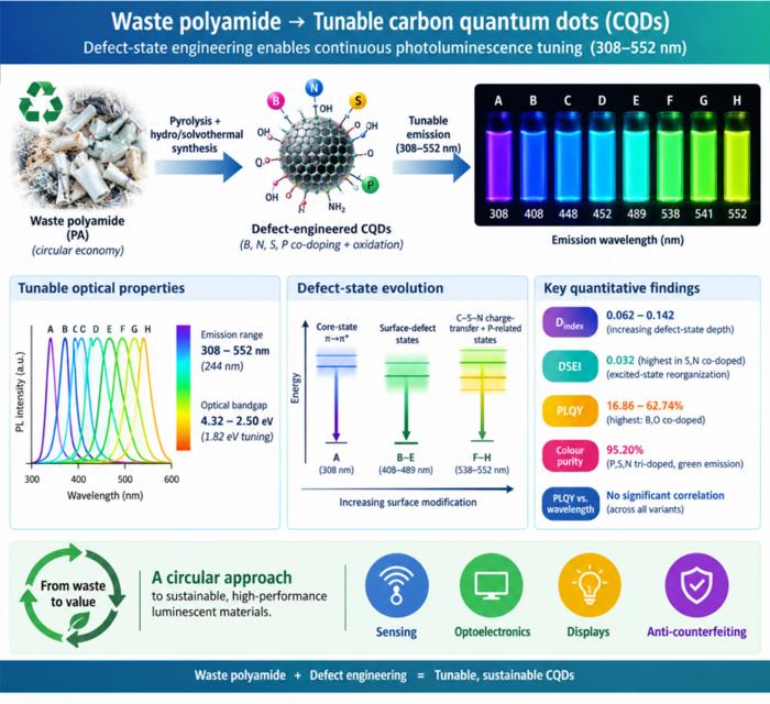
Waste polyamide-derived CQDs show continuous photoluminescence tuning from 308 to 552 nm through sequential defect-state engineering, with optical transition energies decreasing from 4.32 to 2.50 eV. @Christian Ebere Enyoh from Saitama University Carbon quantum dots (CQDs) are fluorescent carbon nanomaterials with potential applications in sensing, optoelectronics, displays, anti-counterfeiting, and environmental technologies. Their optical properties can be adjusted by modifying the carbon...
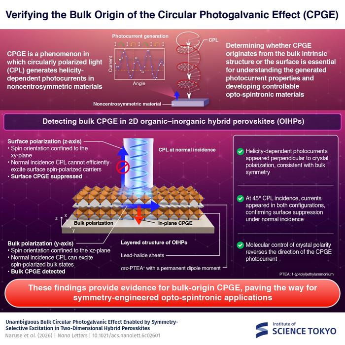
The researchers used circularly polarized light at normal incidence and found that helicity-dependent photocurrents appeared perpendicular to the crystal’s polarization but vanished when measured parallel to it, supporting a bulk origin of CPGE. @Institute of Science Tokyo The circular photogalvanic effect (CPGE), a phenomenon that generates helicity-dependent photocurrents in noncentrosymmetric materials, can originate purely from a crystal's internal structure without contribution from the...
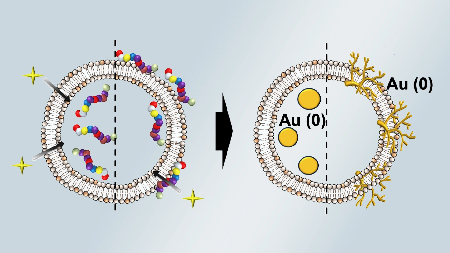
Summary of gold nanoparticle growth programming in liposome based on localization of biomineralization peptide The position of biomineralization peptides within liposomes can influence how gold nanoparticles grow, reports a research team from Institute of Science Tokyo. Peptides localized at the membrane interface promote branched structures, while those confined to the liposome interior favor spherical nanoparticles. The findings offer a new strategy for controlling nanoscale reaction...