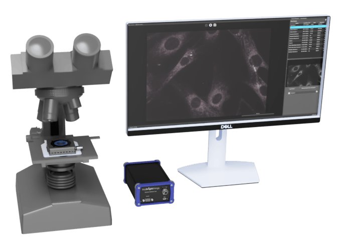
An innovative microscopy technique that enables visualization of features across a wide size range—from nanometers to millimeters—is the HaloMicroscopy system developed by Tiny Bright Things. This system employs Resonance Imaging Microscopy to deliver visible light via an Evanescent Wave (EW) to illuminate samples, revealing details for both sub-visible and visible size ranges. The HaloMicroscopy is a true plug-and-play microscopy system, excellent for researchers needing rapid, label-free, quantitative imaging without complex optical setups. Its simplicity and minimal learning curve make it ideal for both academic labs and biotech companies. HaloMicroscopy is a cutting-edge, snapshot-based technique that delivers full field of view imaging at millisecond speeds. It’s ideal for applications where traditional point-scanning or slow serial acquisition is too limited to capture fast, complex dynamics. HaloMicroscopy is a low-cost, high-speed imaging solution that drastically reduces both equipment cost and experiment time. It’s ideal for labs needing fast, scalable, and accessible imaging for basic analysis — especially in education, diagnostics, or screening environments. Stained and unstained serial sections from liver with confirmed diagnosis of haemochromatosis (abnormal accumulation of iron). The presence of ferric iron granules was confirmed by Perl’s Prussian blue staining. Samples courtesy of Dr Chris Freelance, Melbourne Histology Platform HaloMicroscopy provides data in its most raw, unprocessed form — pixel-level — giving you full flexibility and control over your own data analysis. It’s perfect for those who want to apply custom algorithms or explore subtle biological phenomena without the bias of pre-processed imaging. HaloMicroscopy can characterise samples in almost any state (samples can be run as-is, even at high concentrations or diluted). Unlike conventional microscopy, it allows observation of non-conductive, hydrated or sensitive materials in their natural state-without the need for hydration, coating or extensive processing. This makes it especially useful for studying biological, organic and soft materials. See features across wide range of sizes. Set up and ready to use in under an hour. Capture everything quickly in one shot. Function, not just form Observe real-time biological and material processes. Structure, not just surface Reveal interfaces or internal structure in 3-D. Snapshot of live sample Real-time capture of a living specimen. No Stain, No Label. All the Details. Find things quickly and spend less time finding stuff. Raw and Processed images based on user’s preference
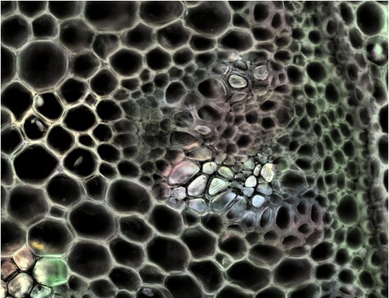
An innovative microscopy technique that enables visualization of features across a wide size range—from nanometers to millimeters—is the HaloMicroscopy system developed by Tiny Bright Things. This system employs Resonance Imaging Microscopy to deliver visible light via an Evanescent Wave (EW) to illuminate samples, revealing details for both sub-visible and visible size ranges. The HaloMicroscopy is a true plug-and-play microscopy system, excellent for researchers needing rapid, label-free, quantitative imaging without complex optical setups. Its simplicity and minimal learning curve make it ideal for both academic labs and biotech companies. HaloMicroscopy is a cutting-edge, snapshot-based technique that delivers full field of view imaging at millisecond speeds. It’s ideal for applications where traditional point-scanning or slow serial acquisition is too limited to capture fast, complex dynamics. HaloMicroscopy is a low-cost, high-speed imaging solution that drastically reduces both equipment cost and experiment time. It’s ideal for labs needing fast, scalable, and accessible imaging for basic analysis — especially in education, diagnostics, or screening environments. Natural and non-destructive characterisation of biological and biomedical samples - Stained and unstained serial sections from liver with confirmed diagnosis of haemochromatosis (abnormal accumulation of iron). The presence of ferric iron granules was confirmed by Perl’s Prussian blue staining. Samples courtesy of Dr Chris Freelance, Melbourne Histology Platform. HaloMicroscopy provides data in its most raw, unprocessed form — pixel-level — giving you full flexibility and control over your own data analysis. It’s perfect for those who want to apply custom algorithms or explore subtle biological phenomena without the bias of pre-processed imaging. HaloMicroscopy can characterise samples in almost any state (samples can be run as-is, even at high concentrations or diluted). Unlike conventional microscopy, it allows observation of non-conductive, hydrated or sensitive materials in their natural state-without the need for hydration, coating or extensive processing. This makes it especially useful for studying biological, organic and soft materials.
CATEGORY
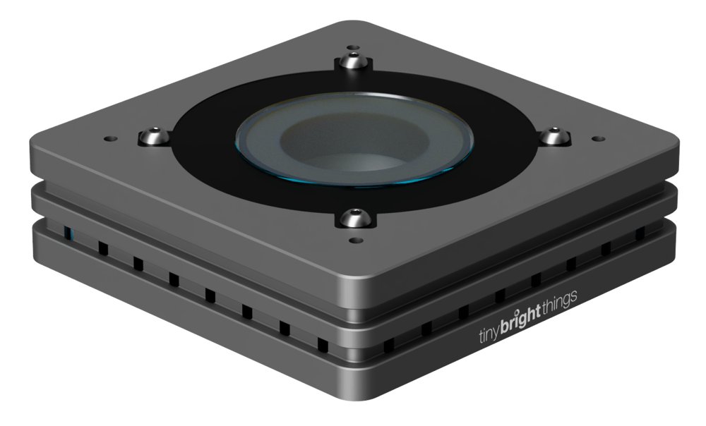
Introducing HaloMicroscopy What is Resonance Imaging Microscopy? The HaloElement allows users to detect and image particles, materials, cells, bacteria, crystals, capsules, histology sections. It can also image opaque samples such as semiconductors, fibres, polymer films, bank-notes and metal particles, in air or in liquid. Key features of the HaloElement include: - Laser free and non-destructive process; - No labels, dyes or stains are required; - Size range 10’s of nanometres up to millimetre scale; - Acquires images as fast as you can take a picture; - Simple sample handling, using a glass slide, cover slip with liquids, in air, or a petri dish; - Integrates with off the shelf heating stages and flow cells for live-cell imaging; - The resultant data and image is owned by you. Underpinned by the discovery of Resonance Imaging Microscopy (RIM) at the University of Melbourne, the HaloElement employs this patented technology to create an Evanescent Wave (EW), used to illuminate samples from the side rather than vertically. A ring on LEDs surrounding the sample is used to generate an EW via total internal reflection at the solid-air or solid-liquid interface. When at least a portion of the sample interacts with the EW, the EW scatters or resonates within the object. The resultant light, scattered by the sample, is detected by conventional light microscopy methods and a composite image is created.
CATEGORY
