By Linkam Scientific Instruments
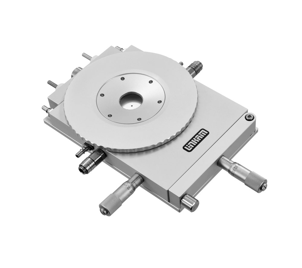
Researchers studied fluid inclusions in quartz samples from an epithermal bonanza-type ore deposit in Mexico using Linkam’s THMSG600. Scientists in China used a Linkam THMSG600 to enable their research into climate change.
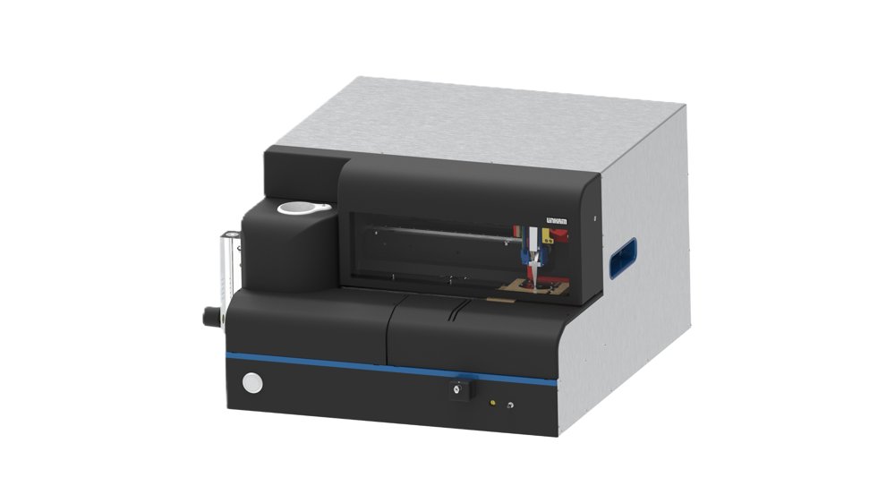
The next generation Cryo Plunger Imaging of biological samples embedded in vitrified ice has become of great interest in recent years as it provides several advantages: the biological sample is in a fully hydrated state with superior preservation down to ultra-structural level, a vitrified sample is naturally compatible with the vacuum required for EM / Single Particle (SPT) / CLEM (Correlative Light and Electron Microscopy) and cryo-fluorescence provides very low photo-bleaching and high signal to noise imaging. Preparation and handling of vitrified samples normally requires special skills and techniques. The novel design of the Linkam CryoGenium makes this a simple and reproducible process. There are several common issues in a conventional plunge-freezing setup. Typically the sample is prepared on an electron microscopy grid and excess liquid is removed inside a humidity chamber using blotting paper to control the ice thickness of the frozen sample. The blotting paper usually needs alignment and blotting force calibration, while the paper does not work consistently over time because it soaks up moisture in the humidity chamber and prevents real-time optical monitoring during blotting. The novel design replaces the common blotting mechanism to improve process stability. The system introduces real-time optical observation of the sample with control of the liquid film thickness. Film thickness is controlled by adjusting the speed at which the sample is drawn from the fluid as well as thinning of the film thickness by controlled suction. When the correct conditions are confirmed by the optical system, the user triggers plunging into a temperature-controlled bath of liquid ethane. Plunging is followed by automated loading into either a special cryo-holder or a cryo-transfer container. Fully automated, programmed operation is also possible. The CryoGenium provides a platform that increases the success rate and quality of plunged grids, enabling improved efficiency and less time wasted at the TEM, resulting in cost savings. Features / Highlights of CryoGenium - Fully Automated System: Automated grid preparation, sample loading, sample grid handling, and cryo-transfer in one compact unit - Real-Time Optical Monitoring: Thin film interference enables water thickness to be monitored prior to plunging - Novel system for control of film thickness: Water film thickness is accurately controlled by precisely controlled suction process - Integrated Glow Discharge: Fully controllable glow discharge unit integrated into the system, with oil-free vacuum pump - Temperature Controlled Ethane Bath: Temperature control of the Ethane bath ensures there is no solidification of the Ethane - Controlled Dew-Point of sample substrate: Dew-point control of sample support substrate inside the humidity chamber allows good stability and control of very thin and delicate liquid films on the sample support - Controlled Temperature sample chamber: The temperature of the sample chamber can be controlled from 4°C to 40°C to ensure optimum conditions for the sample - Autoloading: Automated loading of the grid into cryo-transfer shuttle or cryo-storage box The CryoGenium also features streamlined and easy-to-use software where the workflow, settings and operation of the plunger can be controlled from one place. CryoGenium integrates into your workflow Single Particle Tomography (SPT) SPT is growing rapidly and allows the determination of the molecular structure of proteins using EM and computational classification and reconstruction. However, the sample quality and in particular the ice thickness have major influence on the signal-to-noise of the data. This is why the CryoGenium can become a key tool for SPT because it gives the user better control of ice thickness and sample preparation. Cells, vesicles, and more There is support for cartridges in the CryoGenium where sample grids can be seeded with cells, vesicles etc. and placed in an incubator. The whole cartridge is then placed into CryoGenium and the grids are picked up automatically and processed. FIB-SEM and lamella preparation for cryo-EM tomography Cells can be prepared, plunged, loaded into compatible cartridges for fluorescence or light microscopy screening in the Linkam CMS196 cryo-stage and then transferred to FIB/SEM to prepare lamellas. The CryoGenium supports the CMS196 EM grid cassette, providing the ability to automatically place the plunged grid into the cassette and enabling a seamless workflow from bare grid to vitrified grid ready to be placed on the CMS196 for Cryo-CLEM work, or directly into the Cryo EM.
CATEGORY
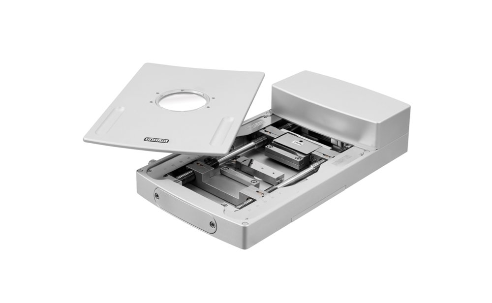
Mechanical sample characterisation with temperature and environmental control modules MFS stage shown above. Understanding the micro- and thermo- mechanical properties of materials is increasingly important. Existing materials are being deployed in new environments and exotic new materials are being developed to meet ever higher demands. Advanced materials such as carbon fibre and graphene are becoming materials of choice for high-end applications such as bike frames, cars, surgical implants and aircraft wings. New materials are being developed to replace or support human body parts such as artificial hips and artificial corneas. While these new materials have many unique and exciting properties, allowing engineers to design and make things that were not possible previously, It is still vital that the physical properties of these materials are characterized and any failure modes well understood. The Modular Force Stage (MFS) is a modular system designed to characterise the mechanical properties of your samples. The system is a new and improved version of the popular TST350 tensile stage with increased sensitivity, resolution and a modular concept. The modular design allows users to have an additional level of control over their experiments with the ability to change grips, heater type and force ranges. The additional options will accommodate a wide range of different samples and applications and be able to test tension, compression and multi-point bending. The MFS can be used with reflected or transmitted illumination as well as other techniques such as X-ray, Raman and FTIR. Its sample chamber is sealed and can be gas purged via the built in valves. There are also options for humidity control and electrical contact posts. MFS on Imaging station, with a sample under tension across the 350°C heater block. Features - Compression and Tensile Force: Test the compressive and tensile properties of your sample relative to temperature and capture high resolution images of the structural changes. - Multi-point bending measurements - Wide range of control parameters: Speed of jaws, distance moved and the force applied can be varied relative to temperature. - Interchangeable Heaters & Grips: The temperature can be controlled from <–195°C up to 600°C depending on heater attached. Heaters and grips can be changed to accommodate a wide range of sample types. - Electrical connections: Optional electrical connections enable electrical measurement on the sample during tensile testing. - Humidity: Add the RHGen humidity unit to your system to accurately control the relative humidity around your samples. - Encoded Distance Measurement: Built in high resolution encoder ensures precise measurement of changes in length. Graphical Data Analyse the mechanical properties of your samples with NEXUS software. Combine it with the NEXUS imaging module to visualise samples throughout your experiment. Real-time graphs are displayed as the experiment is run. You can visualise your data in both temperature-time and force–extension graphs. MFS on Imaging Station
CATEGORY
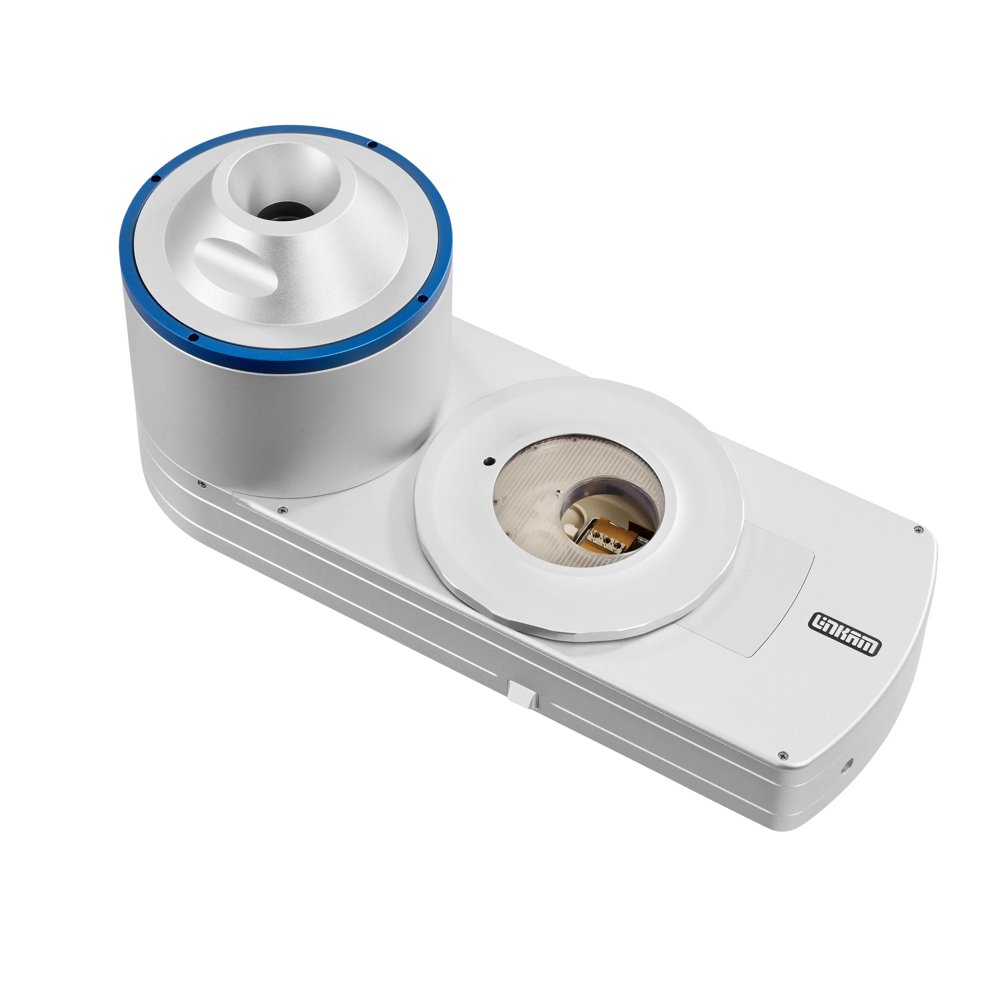
Cryo-Correlative Microscopy Stage CMS196V⁴ Cryo-CLEM stage overview Linkam’s new CMS196V⁴ is a market-leading cryo-correlative microscopy system enabling cryo-fluorescence, the full workflow of Correlative Light and Electron Microscopy (CLEM) and Cryo-super resolution. The CMS196V⁴ maintains the vitrified state of the sample by liquid nitrogen cooling and provides proven capabilities to safely handle and transfer your cryo-samples. These can then be imaged with optical microscopy whilst ensuring they remain free of contamination. The integrated, encoded and motorised XY stage enables the co-ordinate mapping required to find the same sample feature again on complementing modalities, for example TEM, FIB-SEM or X-ray systems. Samples are typically mounted on EM grids or other carriers compatible with electron microscopy, allowing correlated light and electron microscopy. The CMS196V⁴ enables high resolution maps of the grid to be produced quickly and easily in combination with our NEXUS software. Up to 3 grids can be loaded into a specially designed cassette which is transported from a plunge freezer in a small, sealed container. The container is loaded into a pre-cooled dry sample loading chamber fitted on an upright fluoresence microscope. The cassette is then easily loaded onto the viewing bridge using special manipulation tools. Originally developed in collaboration with Prof. Bram Koster's team at Leiden University Medical Centre (LUMC) in the Netherlands, the CMS196 series is the result of years of intensive research and development. “The correlative stage was developed out of the necessity for a tool that uses fluorescence microscopy to help localise structures of interest in samples for high resolution cryo electron microscopy imaging. Over the years the system has evolved into an effective and efficient instrument that enables the imaging of cryo electron microscope images after light microscopy characterisation routinely.” Prof. A.J. Koster - LUMC CMS196V4 Key Features - Self-contained cryo sample observation system with automatic LN2 top-up of the sample chamber which ensures that the sample remains vitrified at liquid nitrogen temperatures (<-195°C) and does not become contaminated. - Short startup time of only 10 minutes with no mechanical connection. - Provides a highly stable platform allowing high precision automated mapping of the complete EM grid with 1 μm resolution. - Self-aligning magnetic sample cassette system ensures safe sample loading, transfer and storage. One cassette can hold up to 3 EM-grids. Dedicated Cassettes support standard EM grids, Autogrids/Clipped Grids, Planchettes, Bessy, membrane carriers (Leica HPF system), Si3N4 frames, CryoCapCell, JEOL CRYO ARM and custom sample holders. - Sample cassette holder for safe and contamination-free sample loading, storage and transfer. - Interchangeable cryo-bridge which allows multiple sample interface options and accommodates different custom needs. - Cordless, heated frost-free lid allows convenient viewing of the sample area. - Integrated LED condenser allows brightfield transmitted light, ideal for initial viewing of the grid and locating the sample. - Integrated sensors for real-time sample temperature monitoring. - The CMS196V⁴ can be configured and operated from the connected joystick touch panel. The joystick wheels allow intuitive sample navigation and swift sample review without a PC. The stage can also be controlled via USB and is supported by multiple software packages. - Support for EM grids, Autogrids (FEI, sometimes referred to as ‘clipped grids’), membrane carriers (Leica HPF system), Bessy grids, Planchettes, CryoCapCell and custom sample holders. - Sample cassette holder for safe and contamination-free sample loading, storage and transfer. - Motorised sample positioning can be operated from the connected joystick touch panel without a PC. The stage can also be controlled via USB for PC control and is supported by multiple software packages. - Connects to plunge-freezing and high-pressure freezing sample preparation protocols. - Built-in bake-out heating cycle to remove moisture in the sample chamber. - Optional objective lens’ heater enhances superior drift performance. - The CMS196V⁴ is compatible with most research-grade upright microscopes and high NA objectives. A quick-release mount further simplifies sample loading for some microscopes.
CATEGORY
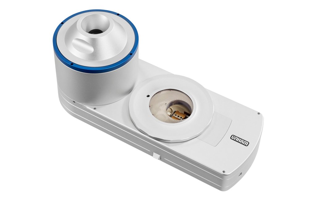
Researchers at The Spanish National Research Council (CSIC), Madrid, Spain used Linkam's CMS196V4 cryo-stage to investigate how growth hormone (GH) changes the metabolism and mitochondrial structure of inflammatory macrophages in their paper, 'Growth hormone remodels the 3D-structure of the mitochondria of inflammatory macrophages and promotes metabolic reprogramming'. Researchers at King's College London in the UK supported the development of early models of our CMS196 V4 stage and continue to test new cryomicroscopy systems including the CryoGenium. Researchers in Barcelona investigate the effect of additives on the effectiveness of cisplatin, a chemotherapy medication, using the CMS196 for cryo-fluorescence miscroscopy. Groen et al. explored how a novel 3D cryo-correlative light and X-ray tomography (CLXT) imaging approach could be used to investigate a promising antifibrotic treatment for myocardial fibrosis, using Linkam's CMS196 to pre-screen vitrified grids. Using a Linkam CMS196 stage Beer et al. demonstrated an innovative, targeted cryoCLEM workflow for tissues, in which cryogenic confocal fluorescence imaging of millimetre-scale volumes is correlated to 3D cryogenic electron imaging directed by a patterned surface generated during high-pressure freezing (HPF). Cryogenic light microscopy is a key mapping step in the correlative imaging workflow. Researchers adapted a standard upright widefield microscope into a cryogenic 3D laser-free confocal system using Linkam's CMS196, to demonstrate the necessary sample preparation steps followed by confocal imaging of biological cells and tissues. Researchers used the Linkam CMS196 for imaging cryopreserved human prostate cancer cells with X-ray tomography. Scientists at the The Institute of Cancer Research in London, UK used Linkam's CMS196 to image mammalian cells with cryo-CLXM microscopy. Researchers used Linkam's CMS196 as part of a Confocal Cryo Fluorescence Microscopy setup to image vitrified biological samples.
CATEGORY
