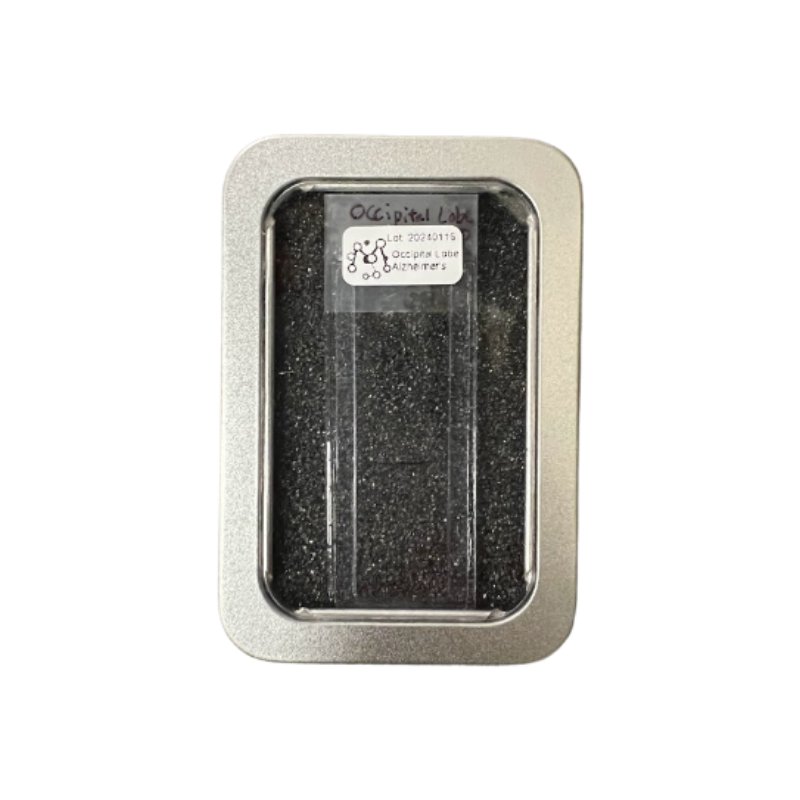
The Magnify Expansion mounted Slide is designed for immediate imaging, eliminating the need for any sample preparation processes. It is ideal for microscopic demonstrations or as a first step in trying Magnify Expansion Microscopy; it helps users evaluate the capabilities of Magnify Expansion microscopy and become familiar with imaging expanded samples. This pre-made slide is also perfect for teaching Expansion Microscopy imaging techniques. It is compatible with a wide range of imaging systems, including slide scanners, widefield fluorescence microscopes, and confocal microscopes in both upright and inverted configurations. The slide works well with dry lenses, water immersion lenses, and oil/silicone immersion lenses of any magnification, offering 3.5x to 11x expansion according to your needs. Each slide comes pre-stained with morphological or customized markers of your choice, ensuring that you can achieve optimal imaging results right out of the box. Slide format Empower super-resolution imaging, achieving CLEM-like quality with <10 nm resolution This Magnify image was captured using a ZEISS Lattice Structured Illumination Microscopy (SIM) 5 with a 63× oil immersion objective (NA 1.4). The sample, an expanded human FFPE kidney tissue, was stained with ACTN4 (magenta) to highlight the tertiary podocyte foot processes. Magnify, combined with the SIM 5’s original 65 nm resolution, enhanced the imaging down to 6-10 nm, achieving clarity similar to Correlative Light and Electron Microscopy (CLEM) but with much simpler and quicker preparation. This ultra-high-resolution capability is invaluable not only for renal pathology but also for a wide range of biomedical research areas, such as neurodegenerative diseases, cancer, and cardiovascular conditions. By providing detailed views of subcellular structures quickly and efficiently, Magnify empowers researchers with SIM scopes to explore complex biological processes, accelerating discoveries and advancing the development of more effective treatments. The Magnify Expansion Slide is ready for imaging with any microscope right out of the box. This dataset was captured using Cephla’s SQUID, a system designed to eliminate barriers to high-end imaging, equipped with a CrestOptics X-Light V2 confocal spinning disk and IMX571 camera, paired with a 63×/1.2 water immersion objective. The mouse brain sample was stained with DAPI, synaptophysin (green), homer (red), and LEL (white), and imaged in high resolution to reveal the detailed structure of neuronal and synaptic components. Magnify’s integration with accessible, high-throughput imaging systems like Cephla’s SQUID enables detailed visualization of synaptic structures without the need for bespoke setups. This approach enhances our ability to study synaptic organization and connectivity in the brain, providing critical insights into neuronal circuitry. By making advanced imaging more accessible, Magnify facilitates broader adoption of cutting-edge research techniques, driving discoveries in brain function and neurological disorders. Rapidly growing catalog of Magnify Expansion slides Example Widefield Fluorescence Image Example Confocol Fluorescence Images
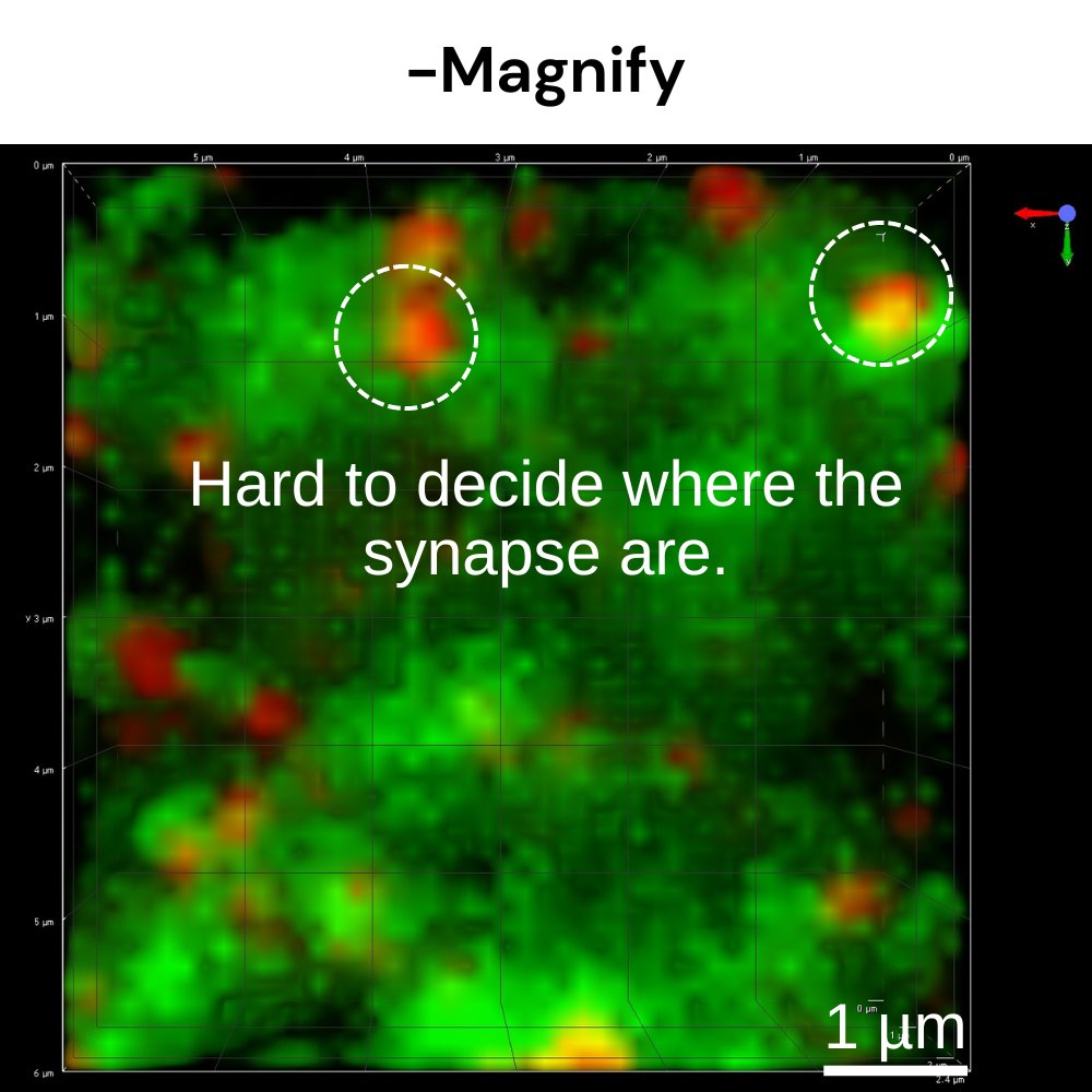
A fast, easy-to-use, and versatile kit that is compatible with any microscope and accessible to everyone. Kit Workflow Summary Magnify™ Kit: Step-by-Step Video Tutorials Part 1 Magnify™ Kit Overview on Freshly Preserved Tissue Part 2 Magnify™ Kit Preparation Step on Freshly Preserved Tissue Part3 Magnify™ Kit Gelling Step on Freshly Preserved Tissue Part 4 Magnify™ Kit Homogenization Step on Freshly Preserved Tissue Part 5 Magnify™ Kit Washing and Staining Step on Freshly Preserved Tissue Part 6 Magnify™ Kit Expansion Step on Freshly Preserved Tissue Magnify™ Kit on Culture Cells Magnify™ Kit Homogenization with pressure cooker on FFPE Unlock potential in any microscope, including low-cost, open source or DIY systems. These images compare mouse brain tissue captured with and without Magnify processing using Cephla's SQUID system (https://cephla.com/). Both samples were stained with DAPI, synaptophysin (green), homer (red), and LEL (white), and imaged using a CrestOptics Cicero confocal spinning disk module, IMX571 camera, and a 63×/1.2 water immersion objective. The integration of Magnify enhances the imaging resolution and clarity, revealing finer details of neuronal and synaptic structures. The application of Magnify significantly improves visualization of critical synaptic proteins such as synaptophysin and homer, providing a more detailed and comprehensive view of synaptic organization and neural connectivity. This enhanced imaging facilitates deeper insights into brain function and the underlying mechanisms of neurological disorders. By combining Magnify with accessible and high-throughput systems like Cephla's SQUID, researchers can achieve superior image quality without reliance on specialized or bespoke microscopy setups, accelerating advances in neuroscience research. Achieve optical ultrastructural contrast with molecular specificity for subcellular localization study This Magnify image was captured using a Nikon Eclipse Ti2 with a CSU-W1 spinning disk confocal module, Andor v.4.2 Zyla sCMOS camera, and a 40×/1.15 water immersion objective. The sample was stained with Tom20 (magenta) and pan-protein stain (black and white), providing an electron microscopy-like context. Using Magnify's up to 11-fold expansion and achieving resolutions of up to 15 nm, this image precisely localizes Tom20 within the subcellular architecture of mitochondria. This capability highlights Magnify's power to pinpoint the exact subcellular locations of proteins, including potential drug targets, and to illustrate how drugs interact within cells. Such detailed imaging is essential for advancing our understanding of cellular processes and the mechanisms of drug action. Compatible with transgenic animals, such as GFP-expressing neurons (in blue). This Magnify image was captured using a Nikon Eclipse Ti2 with a CSU-W1 spinning disk confocal module, Andor v.4.2 Zyla sCMOS camera, and a 40×/1.15 water immersion objective. The sample, stained with DAPI (white), anti-GFP (blue), synaptophysin (magenta), and PSD95 (green), was rendered in 3D to reveal intricate neuronal architecture. Studying detailed synaptic structures is key to mapping neuronal connections and advancing our understanding of connectomes. Magnify delivers high-resolution 3D imaging of SST neurons, revealing synaptic components like synaptophysin and PSD95. Its compatibility with genetically encoded tags, such as GFP, makes it an ideal tool for exploring brain connectivity and synaptic function, providing valuable insights into neurological health and disease. Achieves rapid tissue clearing and declouding within a day, simplifying labeling and imaging. This Magnify image was captured using a Nikon Eclipse Ti2 with a CSU-W1 spinning disk confocal module, Andor v.4.2 Zyla sCMOS camera, and a 10×/0.3 objective. The sample, a ~85 µm thick mouse colon section stained with pan-protein ATTO647N NHS-ester (magenta), was expanded to enhance both resolution and tissue clarity. Unlike other tissue clearing methods that can take days or even weeks, Magnify achieves effective clearing and expansion within a single day. Traditional PFA-fixed tissues, especially those thicker than 30 microns, are often difficult to image due to high autofluorescence and light scattering. After Magnify expansion, the tissue becomes nearly transparent, allowing for much clearer and more detailed imaging, making it the superior choice for rapid and reliable results. Optimized to achieve ultra-high-resolution imaging of pathogens within even the most challenging, collagen-rich tissues, like keratitis-infected corneas. This µMagnify image was captured using a Nikon Eclipse Ti2 with a CSU-W1 spinning disk confocal module, Andor v.4.2 Zyla sCMOS camera, and a 40×/1.15 water immersion objective. The sample, a human cornea infected with Candida albicans, was post-expansion stained with DAPI (cyan), Tomato lectin LEL (yellow), and NHS-ester (magenta), and rendered in 3D to visualize detailed pathogen-host interactions within the dense collagen matrix of the cornea. Studying these intricate pathogen interactions at the single-cell level is critical for understanding the mechanisms of keratitis. µMagnify provides high-resolution 3D imaging that reveals the distinct morphological features of various pathogens, such as the C. albicans cell wall and immune responses within the corneal stroma. Its ability to expand and clear tough tissues like the human cornea enables a new level of detail in pathogen identification, offering valuable insights for advancing diagnostic methods and therapeutic strategies for sight-threatening infections. Preserve lipids for detailed visualization of extracellular vesicles. This Magnify image showcases the detailed ultrastructure of extracellular vesicles within human stem-cell-derived lung organoids. Captured with a Nikon Eclipse Ti2 equipped with a CSU-W1 spinning disk confocal module and Andor v.4.2 Zyla sCMOS camera, the sample was expanded 10.2-fold and stained with Alexa Fluor 488 NHS ester (green) and DiD (magenta). Panel b is 3D reconstruction of the EVs in panel a; Panel c shows a close-up 3D reconstruction of the highlighted EV in a; d is the orthogonal view of c. By achieving resolutions down to 15 nm, Magnify allows for the precise visualization of complex internal structures within extracellular vesicles, which are crucial for understanding their roles in cellular communication, disease progression, and potential therapeutic targeting. Preserving lipids during expansion is key to maintaining the integrity of these vesicles, providing a clear view of their morphology and function. Such insights are essential for advancing our knowledge of how extracellular vesicles contribute to both normal physiology and pathological conditions. Preserve sugars for ultrastructural visualization of blood vessels. These images compare tissue samples captured with and without Magnify processing using the same Cephla's SQUID system setup, equipped with a CrestOptics X-Light V2 confocal spinning disk, IMX571 camera, and a 63×/1.2 water immersion objective. The samples were stained differently to highlight nuclear and vascular structures, with Magnify enhancing the resolution and clarity, making abnormalities more discernible. Magnify's enhanced imaging capability allows for the clear visualization of nuclear abnormalities, which are often linked to cancer, and provides critical insights into vascular pathologies such as tumor angiogenesis. By revealing these subtle yet significant details, Magnify aids in the early detection and study of cancerous changes and vascular developments, offering valuable tools for advancing cancer research and therapeutic strategies.
CATEGORY
A fast, easy-to-use, and versatile kit that is compatible with any microscope and accessible to everyone. Kit Workflow Summary Magnify™ Kit: Step-by-Step Video Tutorials Part 1 Magnify™ Kit Overview on Freshly Preserved Tissue Part 2 Magnify™ Kit Preparation Step on Freshly Preserved Tissue Part3 Magnify™ Kit Gelling Step on Freshly Preserved Tissue Part 4 Magnify™ Kit Homogenization Step on Freshly Preserved Tissue Part 5 Magnify™ Kit Washing and Staining Step on Freshly Preserved Tissue Part 6 Magnify™ Kit Expansion Step on Freshly Preserved Tissue Magnify™ Kit on Culture Cells Magnify™ Kit Homogenization with pressure cooker on FFPE Unlock potential in any microscope, including low-cost, open source or DIY systems. Achieve optical ultrastructural contrast with molecular specificity for subcellular localization study Compatible with transgenic animals, such as GFP-expressing neurons (in blue). Achieves rapid tissue clearing and declouding within a day, simplifying labeling and imaging. Optimized to achieve ultra-high-resolution imaging of pathogens within even the most challenging, collagen-rich tissues, like keratitis-infected corneas. Preserve lipids for detailed visualization of extracellular vesicles. Preserve sugars for ultrastructural visualization of blood vessels.
CATEGORY
