By Nt-mdt
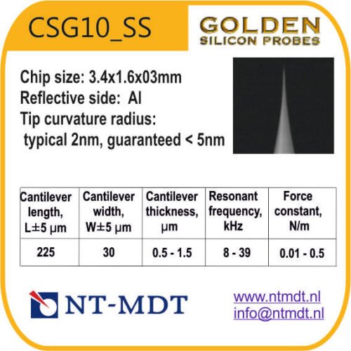
€850,00 Super Sharp High Resolution Silicon AFM Cantilevers CSG10_SS series are designed for Contact applications. Typical Resonant Frequency 22 kHz (guaranteed range 8-39kHz), Typical Force Constant 0,11 N/m (guaranteed range 0,01-0,5N/m).Typical tip curvature radius is 2nm. Using the probes with sharpened tips you will be able to increase resolution of the obtained images especially on objects with typical sizes of several nanometers.Cantilever has Al reflective side coating to increase laser signal. Probes are packed in boxes with 5 pieces. General Features Material Single Crystal Silicon, N-type, 0.01-0.025 Ohm-cm, Antimony doped Chip size 3.4×1.6×0.3mm Reflective side coating Al Tip coating No Tip curvature radius typical 2nm, guaranteed 5nm Special Features Cantilever series: CSG10_SS Cantilever length, L±10µm: 225 Cantilever width, W±5µm: 30 Cantilever thickness, T±0.5µm: 1.0 Resonant frequency, kHz: min 8, typical 22, max 39 Force constant, N/m: min 0.01, typical 0.11, max 0.5
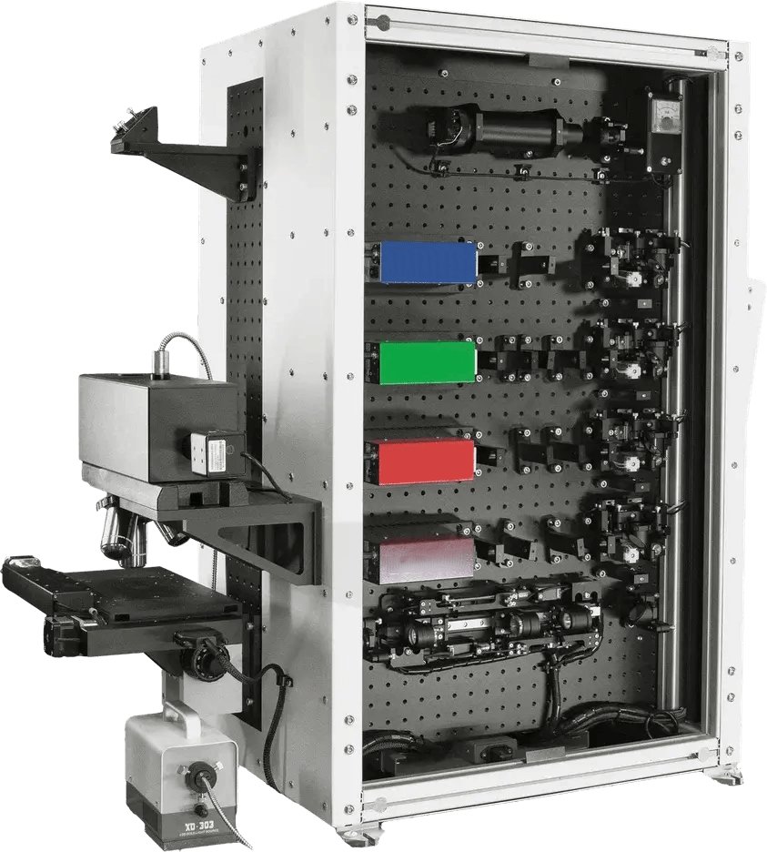
RAMAN AND PHOTOLUMINESCENCE CONFOCAL MICROSPECTROMETER. COMPREHENSIVE PHYSICAL & CHEMICAL MICROANALYSIS. APPLICATIONS - In pharmacy to get tablet surface chemical images, to visualize the distribution of components, including depth profiling and optical sectioning. - For rapid analysis of raw materials quality and authenticity, including working through transparent packaging without opening it, since polyethylene and other transparent polymers, as well as glass, have a very small Raman scattering cross-section. - In a chemical laboratory, polymer synthesis and monomers conversion can be monitored in real time by spectral signal (spectrometers are equipped with the special optical fiber probes to control directly the reaction mixture). - In the semiconductor industry to perform stress and strain mapping of silicon layers, to study the distribution of impurities and contaminants. - Using an inverted optical microscope to make 3D spectral maps of cells, including measurements in vivo, to study lipids, proteins, amino acids. - In nanotechnology to analyze the spectra of nanotubes, graphene layers, and other nanomaterials. - In criminology to perform a rapid non-destructive analysis of the sample chemical composition, e.g., to verify the authenticity of a written document. In archaeology and art history to determine the exhibits authenticity and to identify the artifacts origin, the artistic pigments and the gemstones. Raman spectroscopy is a non-destructive, non-contact and fast method for physical and chemical analysis which does not require any special sample preparation. The method allows to analyze solid, liquid, powdery samples, gases, solutions, suspensions, etc. No vacuum or other special conditions are required for measurements, spectra are usually recorded at atmospheric environment and room temperature. The spectra obtained provide qualitative and quantitative information. Raman peaks positions indicate the chemical compound and its functional groups while the intensity indicates its concentration. An important information is also provided by shifts in peak positions with changes in temperature or pressure. Together with infrared (IR) spectroscopy, Raman spectroscopy joins a group of techniques called vibrational spectroscopy. Both methods of spectroscopies study the same vibrational levels of molecules, but in different ways. A Raman spectrometer integrated with an optical microscope (including a confocal one) is called “Raman microscope” or “Raman microspectrometer”; it has a number of important advantages. The excitation laser is focused into a small region near the microscope objective lens focus determined by the emission wavelength, the spectral Raman signal is emitted from the same region. It provides localized measurements with the possibility for the spectra being referred to various points on the sample surface. Confocal Raman microspectroscopy with a small aperture provides high spatial resolution (sub-μm) and 3D mapping possibility. An optically transparent 3D sample can be studied layer by layer, and each thin layer of the sample can be explored separately with no need to make cross-sections and to destroy the sample. The LS RamBo 620 microspectrometer is equipped with a high-precision motorized scanning stage and/ or motorized scanning mirrors for 2D/ 3D point-to-point mapping of spectra from a given area/ volume of the sample (Raman mapping). In Raman spectroscopy it is possible to use low-power excitation lasers without a risk of the sample heating, the sample destruction, and the appearance of the thermal radiation parasitic lines. The spectra quality is unaffected by the atmospheric air, including the presence of water, so there is no need to dry or vacuumize the cuvette compartment. The spectra of aqueous solutions, including organic compounds, can be quantitatively analyzed without an uncontrollable absorption of the signal by an aqueous environment. The LS RamBo 620 spectrometer has the modular structure. A wide choice of excitation lasers and optical microscopes, automatic switching between excitation laser channels, automatic optical and mechanical parts adjustment to achieve maximum efficiency – all this makes the system flexible and solves a huge number of research tasks. Stokes shifts from 100 cm-1 (optionally from 50 cm-1) can be measured. The monochromator measurement error limit by wavelength is 1 Å (grating 600 gr/mm). The instrument is controlled by the LS RamBo software. To work on the spectrometer, one may use various commercially available laser sources (with “free space” or fiber input type) and detectors (multi-channel or single-channel). Simplified optical scheme of the LS RamBo 620 operation - L1, L2, L3, L4 – built-in lasers - L5 – external laser - BE1, BE2, BE3, BE4 – beam expanders - LL1, LL2, LL3, LL4 – laser line cleaning filters - ND1, ND2 – neutral density filter wheels - SH1, SH2 (optional) – shutters - BS – beam splitter unit (edge filters) - A1 – analyzer - P1 – polarizer - HWP – half-wave plates wheel - 1, 2, 3, 4, 5, 13 – movable mirrors - 10, 11, 12 – mirrors - 6 (optional) – semi-transparent plate - 7 (optional), 15 – focusing optics - 8 (optional), 14 – motorized mirrors - 9 (optional) – slit - 16 – confocal aperture - 17, 19 – concave mirrors - 18 – diffraction grating unit (3 or 4 gratings on rotating turret) - 20 – calibration lamp - 21 (optional) – movable mirror - 22 (optional) – second spectral detector - PMT (optional) – photomultiplier tube THE COLORS ON THE SCHEME - Laser radiation from source. - Laser radiation scattered by the sample. - Long-wavelength (Stokes) radiation component emitted by the sample. To realize Raman confocal measurements. - Short-wavelength (anti-Stokes) radiation component emitted by the sample + laser radiation reflected from the sample. The laser line and the short-wavelength (anti-Stokes) radiation component intensities are quite different, so we assume the intensity of the reflected laser radiation is detected only, and the laser confocal measurement mode is realized. LS RamBo 620 front side with doors removed LS RamBo 620 back side with doors removed TECHNICAL DATA Diffraction gratings up to 4 pcs. on the turret, to choose from: 150, 300, 600, 1200, 1800, 2400 gr/mm Laser sources Quantity up to 5 (4 built-in + one external) Power up to 100 mW Wavelength range from 400 to 800 nm Polarization Linear Beam profile Gaussian TEM 00, single longitudinal mode (SLM) Monochromator entrance slit width from 0 to 2000 µm step – 1 µm Edge/notch filters 8-position wheel, for filters Ø 12.5 mm and Ø 25 mm Beam expander optimized to fill the entrance pupil of the lens Calibration lamp two-element hollow cathode Detectors CCD detector CCD PMT (optional) PMT APD (optional)APD KEY FEATURES - Czerny-Turner spectrometer with 3 or 4 motorized diffraction gratings on a turret - Focal length 620 mm - Possibility to install up to 5 excitation lasers, internally or/ and externally - Possibility to install different detectors: - CCD detector to visualize the whole emission spectrum - APD or PMT (including photon counting mode) for weak signals - Motorized polarization optics in the excitation and emission channels - Automated switching between the excitation lines - Spatial resolution limited by the diffraction laws - Motorized confocal aperture for sensitivity and spatial resolution optimization - Beam expanders for complete filling of the pupil lens
CATEGORY
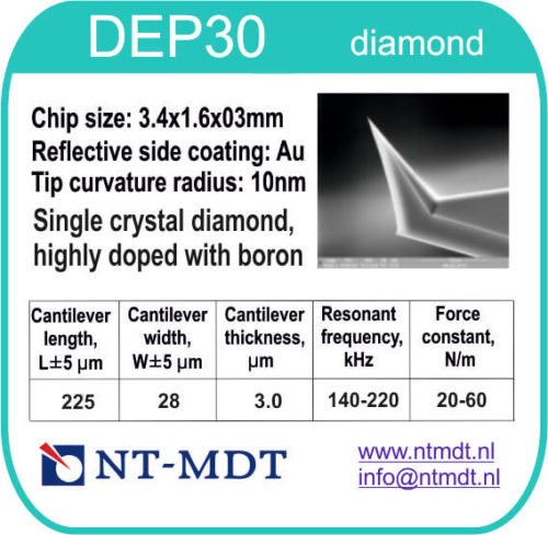
Unique probes with single crystal diamond tip for topography and electrical measurements. Nominal values: force constant - 40 N/m, resonance frequency – 180 kHz. Highly Conductive Long Lasting Sharp Diamond Probes formed by a unique patented process ensure the best possible wear and electrical performance. The tips are sharper and last longer than any other electrical AFM probe. The probes are highly resistant to mechanical destructions and keep their sharpness during the whole working day and more! These probes enable highly repeatable high resolution operation to ensure you consistently get the best possible data from your system. The conductive diamond coating is highly doped with boron which leads to a macroscopic resistivity of 0.003 - 0.005 Ohm•cm. A sharp tip is formed from a single diamond crystal which yields an unsurpassed combination of resolution, mechanical properties and electrical performance. Contact resistance is between 10k and 100k ohms depending on contact radius measured on a silver coated surface. A gold reflex coating is deposited on the detector side of the cantilever to enhance the reflectance of the laser beam. Upon customers' request the next options are available: - super sharp probes with typical curvature radius 2nm; - probes with another lever specification; - probes with individually characterized spring constant and tip radius to enable fully quantitative nanomechanical measurements. Cantilever specifications Geometry: Rectangular Material: Diamond coated Si Cantilever bending: < 3° Bulk tip specifications Si geometry: Rotated (Symmetric) Si tip height (h): 15 ± 2 μm Front angle: 25 ± 2° Back angle: 22 ± 2° Side angle: 18 ± 2° Tip offset: 10 – 20 μm Single crystal diamond tip specifications Geometry: Cone Tip radius: Typical 7nm, guaranteed <10nm Tip height (h): 300 nm ± 100 nm Tilt angle: 0 ± 1° Tip material: Single crystal diamond Highly doped with boron Cone ½ angle: 15 ± 2° General Features Chip size 3.4×1.6×0.3 Reflective side coating Au Tip coating Single crystal diamond, Highly doped with boron Tip curvature radius typical 7nm, guaranteed <10nm Special Features Cantilever series DEP30 Cantilever length, L±10µm: 225 Cantilever width, W±5µm: 28 Cantilever thickness, T±0.5µm: 3.0 Resonant frequency, kHz min 140, typical 180, max 220 Force constant, N/m min 20, typical 40, max 60 Additional information Quantity: Set of 10 items, Set of 30 items
CATEGORY
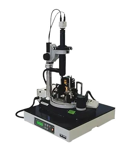
Universal scanning probe microscope, realizing more than 40 methods of atomic force and tunneling microscopy. The functionality of the basic model can be significantly extended; the microscope can be easily integrated with other analytical methods into more powerful measuring systems. APPLICATIONS - Materials science: electrical, magnetic and mechanical properties - Polymers and thin organic films - Biology/ Biotechnology - Nanomaterials/ Nanostructures - Nanomachining/ Nanomanipulation NTEGRA Prima is a multifunctional system designed for the most common tasks in the field of Scanning Probe Microscopy. The device can perform more than 40 measurement techniques, allowing for precise and high-resolution analysis of physical and chemical properties. The microscope is versatile in its functionality and measurement capabilities, yet convenient and easy to use. NTEGRA Prima offers the possibility to examine samples at the atomic and molecular level in different environments – in air, liquid and controlled environment. The modular design provides a unique opportunity to configure the instrument for specific applications and research methods. The NTEGRA Prima offers several scanning options: scanning by sample or scanning by probe, as well as simultaneous probe and sample scanning (so-called DualScan). Scanning by sample is performed using interchangeable scanners integrated into the basic unit and is suitable for small samples and ultra-high resolution. Scanning by probe is performed by scanning heads. The advantage of this type of scanning is a large range (up to 100x100x10μm) and the ability to examine large samples (up to 100mm in diameter). In addition, such measuring heads can be used as stand alone measuring devices, which makes it possible to examine objects of unlimited size. The unique DualScan mode allows to extend the scanning range up to 200x200x20 µm, which can be useful for studying living cells or MEMS components. Built-in three-axis closed-loop control sensors trace the actual displacement of the scanner and compensate for unavoidable imperfections of piezoceramics, such as non-linearity, creep and hysteresis. The sensors have a low noise level, allowing for working with closed-loop controls on very small areas. This is particularly valuable for nanomanipulation/nanolithography operations and for measuring very small objects. The integrated optical video system is ergonomic, easy to use and provides spatial resolution of 1 µm. It allows real-time visualization of the scanning process. The front panel of the instrument has an LCD monitor that displays temperature and relative humidity information while the instrument operation. MEASURING MODES AND TECHNIQUES In air&liquid: AFM (Contact + Semi-Contact + Non-Contact)/ Lateral force microscopy/ Phase Imaging/ Force Modulation/ Adhesion Force Imaging/ Lithography: AFM (Force) In air only: STM/ Magnetic Force Microscopy/ Electrostatic Force Microscopy/ Scanning Capacitance Microscopy/ Kelvin Probe Microscopy/ Spreading Resistance Imaging/ Piezoresponse Force Microscopy/ Lithography: AFM (Current, Voltage) TECHNICAL DATA Scan type / Scanning by sample / Scanning by probe Sample size: Up to 40 mm in diameter, to 15 mm in height / Up to 100 mm in diameter, up to 15 mm in height Sample weight: Up to 100 g / Up to 300 g XY sample positiniong: 5×5 mm / 5×5 mm Positioning resolution: 5 µm / 5 µm Scan range: 100x100x10 µm, Up to 200x200x20 µm (DualScan mode) / 100x100x10 µm, Up to 200x200x20 µm (DualScan mode) Non linearity, XY (with closed loop sensors): ≤0.1% / ≤0.15% Noise level, Z (RMS in bandwidth 1000 Hz): With sensors 0.04 nm (typically), ≤0.06 nm / 0.06 nm (typically), ≤0.07 nm; Without sensors 0.03 nm / 0.05 nm Optical viewing system: Optical resolution 1 µm / 3 µm; Field of view min 0.4-0.3 mm / 0.5-0.4 mm; Field of view max 4.9-3.7 mm / 6.5-4.9 mm; Continuous zoom available / available Vibration isolation: Active 0.6-200 Hz / 0.6-200 Hz; Passive 200 Hz / 200 Hz Scanning head can be configured to serve as a stand-alone device to measure specimens of unlimited sizes. Option. KEY FEATURES Versatile tool for scanning probe microscopy NTEGRA Prima integrates more than 40 techniques commonly used in Scanning Probe Microscopy (SPM), spectroscopy and lithography. Consequently, Prima is an ideal SPM tool for thoroughly investigating the physical and chemical properties of samples with the highest possible precision and performing high-resolution scans. Simultaneous optical control and scalable possibilities An integrated optical viewing system with a resolution of up to 1 µm allows targeting the tip and controlling the measurement process in real time. The NTEGRA Prima system supports all types of scanning required for various samples. It can be used for scanning by sample, by probe or simultaneously by both methods. This also increases the scanning range to up to 200 µm in the XY plane and up to 20 µm along the Z axis. Precise and accurate Prima has built-in closed-loop capacitive sensors for all axes (X, Y, Z). These sensors measure the actual scanner displacement and compensate for parasitic piezoceramics properties including non-linearity, hysteresis and creep with extremely low noise level. This enables closed-loop control on the small fields – just what you need for precise lithography with perfect and accurate linearity. Modular design The NTEGRA platform is designed with an open hardware and software architecture. This allows various compatible assemblies and components to be used in the instrument using a simple plug&play method. Due to the modular organization, the specialization of the basic atomic force microscope can be easily changed or expanded. This design provides the unique ability to configure the instrument for specific applications and specific research techniques. Large selection of optional features Thanks to its open architecture, the functionality of the NTEGRA Prima can be significantly expanded. The modular design allows the system to be supplemented with external magnetic field measurements, sample temperature experiments, near-field optical microscopy, Raman spectroscopy, and many other options.
CATEGORY
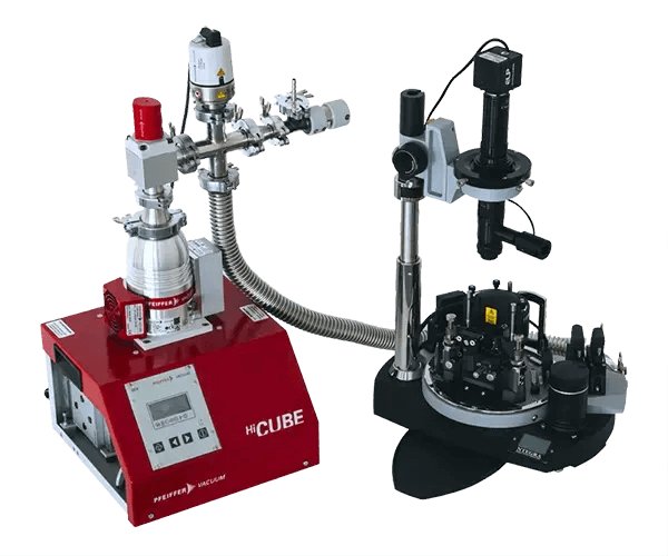
Scanning Probe Microscope for experiments in air, controlled environment and low vacuum. Single- and multi-pass techniques Measurement of weak forces (electrical, magnetic, mechanical, etc.) High sensitivity and measurement accuracy Thorough environment control, low vacuum Open architecture to enhance functionality NTEGRA Aura is a Scanning Probe Microscope for experiments in air, in controlled atmosphere and in low vacuum conditions; ideal for both single- and multi-pass techniques and measurement of weak forces (electrical, magnetic, mechanical, etc.). The Q-factor of the cantilever in vacuum increases, thus gaining the sensitivity, reliability and accuracy of "probe-sample" light forces measurements. At the same time, a shift of pressure from atmospheric one to a 10-2 Torr value provides a tenfold increase in the Q-factor. Further vacuum pumping causes the Q-factor to reach its plateau and changes insignificantly. Therefore, NTEGRA Aura offers the optimal "price/quality" ratio, as compared to high-vacuum devices. It takes only a few minutes to achieve the vacuum required for a tenfold Q-factor increase. At the same time the system is compact, easy to operate and maintain. The NTEGRA Aura has built-in closed-loop control for all the axes, an optical system with 1 um resolution and the ability to work with over 40 different AFM techniques. Due to open architecture, the functionality of the NTEGRA Aura can be extended significantly: to specialized magnetic measurements in an external magnetic field (horizontal, up to +/-2500 Gauss; vertical, up to +/-1100 Gauss, high-temperature experiments (heating to 150 C with temperature maintaining precision of 0.05 C), etc. MEASURING MODES AND TECHNIQUES AFM (Contact + Semi-Contact + Non-Contact)/ Lateral Force Microscopy / Force Modulation Microscopy / Magnetic Force Microscopy/ Electrostatic Force Microscopy / Scanning Capacitance Force Microscopy/ Kelvin Probe Force Microscopy / Piezoresponce Force Microscopy/ Spreading-Resistance Imaging / STM / Lithography: AFM (Force + Voltage + Current), STM APPLICATIONS - Materials science: electrical, magnetic and mechanical properties - Chemically sensitive samples - Polymers and thin organic films - Nanomaterials and nanostructures - Nanomachining/nanomanipulations TECHNICAL DATA Scan type: Scanning by sample / Scanning by probe Sample size: Up to 40 mm in diameter, up to 15 mm in height / Up to 100 mm in diameter, up to 15 mm in height Sample weight: Up to 100 g / Up to 300 g XY sample positiniong: 5x5 mm / 5x5 mm Positioning resolution: 5 um / 5 um Scan range: 100x100x10 um, Up to 200x200x20 um (DualScan mode) / 100x100x10 um, Up to 200x200x20 um (DualScan mode) Non linearity, XY (with closed loop sensors): <= 0.1% / <= 0.15% Noise level, Z (RMS in bandwidth 1000 Hz): With sensors 0.04 nm (typically), <=0.06 nm / 0.06 nm (typically), <=0.07 nm; Without sensors 0.03 nm / 0.05 nm Noise level, XY (RMS in bandwidth 200 Hz): 0.2 nm (typically), <=0.3 nm (XY 100 um) / 0.2 nm (typically), <=0.3 nm (XY 100 um) Optical viewing system: Optical resolution 1 um / 3 um; Field of view min 0.4x0.3 mm / 0.5-0.4 mm; Field of view max 4.9x3.7 mm / 6.5x4.9 mm; Continuous zoom available / available Environment control: air humidity control, gas atmosphere, vacuum measurement / air humidity control, gas atmosphere, vacuum measurement Vacuum system: Pressure 10-2 Torr / 10-2 Torr Electrochemical measurements: available / available Vibration isolation: Active 0.7-1000 Hz / 0.7-1000 Hz; Passive above 1 kHz / above 1 kHz The scanning head can be used as a stand-alone device for measuring specimens of any sizes Option KEY FEATURES Experiments in a controlled environment NTEGRA Aura is intended for SPM experiments in low vacuum and in a controlled environment. Multifunctionality NTEGRA Aura realizes more than 40 Scanning Probe Microscopy (SPM) techniques, including spectroscopy and lithography. It is an ideal tool for investigating physical and chemical properties of samples thoroughly with the highest possible precision and performing high-resolution scans. Light forces measurements In vacuum, the cantilever Q-factor increases, thus the sensitivity, accuracy and reliability of the light force measurements between the probe and the sample also increase. It is especially important for noncontact modes, such as Magnetic Force Microscopy, Electrostatic Force Microscopy and Scanning Capacitance Microscopy. Switching from atmospheric pressure to a 10-2 Torr vacuum provides a ten-fold increase in the Q-factor. Precise and accurate Prima has built-in closed-loop capacitive sensors for all axes (X, Y, Z). These sensors measure the actual scanner displacement and compensate for parasitic piezoceramics properties including non-linearity, hysteresis and creep with extremely low noise level. This enables closed-loop control on the small fields - just what you need for precise lithography with perfect and accurate linearity. Modular design The NTEGRA is designed with an open hardware and software architecture. This allows various compatible assemblies and components to be used in the instrument using a simple plug&play method. Due to the modular organization, the specialization of the basic atomic force microscope can be easily changed or expanded. This design provides the unique ability to configure the instrument for specific applications and specific research techniques. Large selection of optional features Due to its open architecture, the functionality of the NTEGRA Aura can be significantly expanded. The modular design allows the system to be supplemented with external magnetic field measurements, sample temperature experiments, near-field optical microscopy, Raman spectroscopy, and many other options.
CATEGORY
