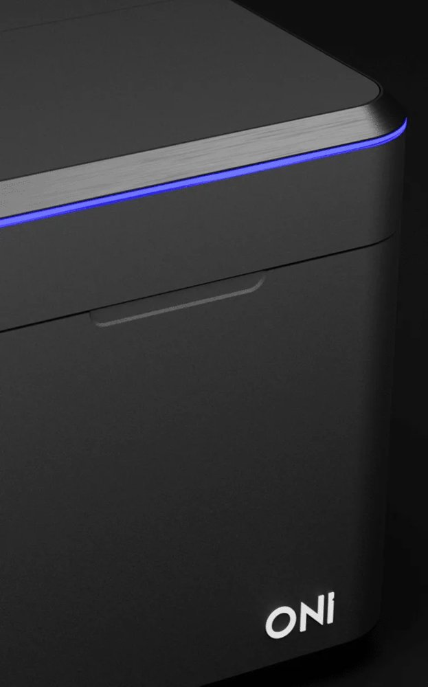
Simple. Engineered. Dynamic. Powerful. A new era in super-resolution imaging Meet Aplo Scope – ONI’s new single-molecule super-resolution microscope designed for effortless molecular imaging. Perfect for researchers of all levels, Aplo Scope combines advanced imaging, precise laser control, and intuitive software to deliver high-quality results, from research on the foundations of life to drug discovery, biomarker research, and disease studies. With its compact design, Aplo Scope transitions seamlessly from live-cell to super-resolution imaging down to 15 nm resolution, without complex setup.Its unparalleled ease of use and integration with the Aplo Platform make it ideal for uncovering molecular interactions and driving breakthroughs. Simple Designed to simplify complex microscopy with one easy-to-use tool. Start imaging quickly, generate data with ease, and unlock answers quicker than ever before. Engineered Robustly engineered for stability, consistency, and reliability, to maximize reproducibility across experiments and users. Dynamic Seamlessly transition between live-cell imaging and super-resolution without reconfiguration for unmatched flexibility in one platform. Powerful Compact footprint, yet powerful enough to enable complex scientific inquiry. Aplo Scope is designed to fit into your lab, not the other way around. The new and groundbreaking SMLM microscope More Channels, More Insights Flexibility Redefined Maximize Throughput Homogeneous Illumination More Channels, More Insights Multicolor imaging reveals biological context Achieve maximal spectral separation with minimized cross-talk and background thanks to an optimized filter set including single, dual, and quad options. Using only 4 lasers, you can generate detailed multi-color panels of 5 or more labels to elucidate complex protein and particle interactions that span both large pan-cellular structures and nanoscale molecular features. Leverage the rapid laser switching with multiband filters for nearly simultaneous multi-channel imaging. Flexibility Redefined Live cell to super-resolution imaging Effortlessly capture dynamic cellular events in real-time, then zoom into molecular details with super-resolution precision — all within a single, intuitive platform designed to enable your cutting-edge science. From low-power live-cell imaging to high-power super-resolution imaging, the Aplo Scope eliminates the need for hardware reconfiguration. Aplo Scope includes unique illumination stability, even at extremely low power (<0.01 kW/cm2), to minimize phototoxicity in extended live cell imaging sessions. At the other end, high laser powers enable rapid, super-resolution imaging to uncover molecular level details. Maximize Throughput Expansive field-of-view Aplo Scope contains a 110 by 110 micrometres field-of-view, in both diffraction-limited and single-molecule localization microscopy. The expansive field-of-view significantly increases the chances of capturing rare events and elusive phenotypes. Detect ultra-rare cell populations and particle biomarkers with unmatched throughput, accelerating groundbreaking discoveries and driving transformative breakthroughs. Homogeneous Illumination Variation that reflects biology Aplo Scope produces a consistently flat, symmetric illumination across the entire field of view, vital for statistical and comparative intensity analysis. With consistent resolution and precision edge to edge, every frame produces more data, maximizing throughput of critical and sensitive assays. This enables quantitative molecular studies to better understand crucial mechanisms in disease and drug discovery studies. Key features of Aplo Scope Next-gen lasers Power density continuously adjustable from 0.01 through 3.325 kW/cm2 Light engine Packed with technology to enable diffraction-limited and SMLM imaging Illumination Illumination angle control from Epi to HILO and TIRF. Consistent symmetric illumination across the field-of-view Temperature control Precise temperature control up to 40°C Filter Wheel Four laser channels can be interlaced with any combination of emission filters to perform multicolor imaging, easily generating 5-color overlays Camera Sensitive next-gen CMOS camera for low noise, high-speed imaging Focus Z-lock Rapid and precise focus with locking technology to minimize Z-drift Field-Of-View 110 by 110 micrometres in both diffraction-limited and single-molecule microscopy Stage Fully automated piezo stage fits most microscopy slides and plates Mechanical stability Integral dual-stage isolation system minimizes vibration Sample enclosure Integral lid interlock ensures user safety, class 1 laser instrument Aplo Scope Aplo Scope is the flagship innovation behind the Aplo Platform: solid performance in a versatile, easy-to-use microscope. Experience a range of imaging modalities with this highly compact microscope designed to simplify multicolor super-resolution microscopy. Aplo Scope allows you to start imaging right away, no dark room or optical table is required. Its integrated software enables fixed or live imaging anywhere, and seamless up to 5 color multimode images! How much training is needed to get started using Aplo Scope? With unparalleled ease-of-use and simplicity, Aplo Scope has been designed to eliminate barriers to entry into the traditionally complex world of super-resolution microscopy and single-molecule imaging, and allow users of any skill level to utilize the instrument within one day of training. What are the dimensions of Aplo Scope? The footprint of the microscope body is as small as a standard laptop 220 mm (w) x 421 mm (d) x 242 mm (h), the light engine measures 215 mm (w) x 420 mm (d) x 456 mm (h) and looks like a computer tower. They’re attached to each other by a 1.5 m long cable and, importantly, can be used on any bench or desk without needing a dark room or optical table. Is Aplo Scope compatible with third-party kits and data analysis tools? Yes, samples can be stained with any super-resolution microscopy-compatible dye and imaged using Aplo Scope. However, this would require manual acquisition and analysis within CODI, or a third party application. ONI offers end-to-end kits where sample preparation, image acquisition, analysis and reporting are all integrated into a workflow, making imaging a seamless and more automated experience.
This kit contains two single-domain antibodies (sdABs) for DNA-PAINT as well as the necessary imager strands and buffers that are required for sample preparation and imaging. Each sdAB can be coupled to a single DNA-PAINT docking site and two sdABs can bind one primary antibody, resulting in two docking sites per primary antibody. The reagents are tested and optimized for the Nanoimager. Simply add your primary antibodies of interest and you are ready for 2 color DNA-PAINT imaging.
CATEGORY
This kit (MASSIVE-sdAB ONI 2-PLEX Kit) includes two single-domain antibodies (sdABs) for DNA-PAINT, along with imager strands and buffers for sample prep and imaging. Each sdAB binds a DNA-PAINT docking site, with two sdABs per primary antibody. Optimized for ONI microscopes—just add your primary antibodies for 2-color DNA-PAINT imaging.
CATEGORY
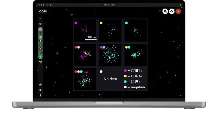
When combined with our newest software offering, AutoEV, your system will be calibrated and optimized to acquire and analyze a 4-lane chip automatically and deliver a comprehensive report of EV size and positivity for each individual lane in 90 mins.
CATEGORY
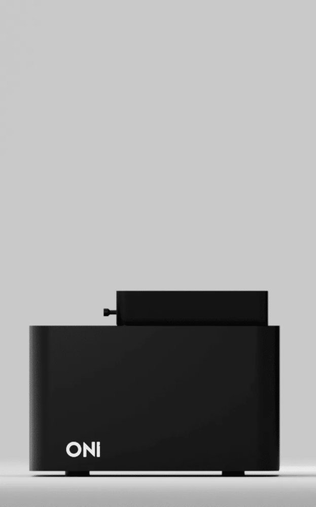
A benchtop super-resolution microscope for your imaging needs Key features Compact A microscope with a smaller footprint than an A4 sheet of paper and a desktop light engine with powerful lasers Robust Super-resolution on your benchtop or biosafety cabinet, no need for an optical table or dark room Easy to use Quick start for imaging, no alignment or calibrations required and real time data build up Flexible Different imaging & illumination modes for either fixed and live cell imaging, and programmable workflows Mechanical stability Designed to be vibration-free, no optical table needed Illumination angle Illumination angle control from 0-65° Interlock pin Securely keeps laser light in, making it a class I instrument Heating Up to 42°C, ideal for for live cell imaging and single-particle tracking Objective Standard 100X NA 1.45. Field of View (FOV): 50 μm x 80 μm Lid Keeps external light out, no need for a dark room Stage Accommodates standard microscopy slides. Travel range: 17.5 x 17.5 x 5 mm XYZ Flat-field homogeneous laser illumination Even illumination across the FOV Multiple lasers 4 lasers, with simultaneous 2 color imaging (dichroic mirror split at 640 nm) Laser power High power ONI lasers Power density at sample: 4 kW/cm². Power at source of up to 1W LED array For brightfield illumination Camera Hamamatsu Orca Flash4.0 v3. Up to 10 ms / 100 Hz full FOV Investigate your sample using different imaging technologies dSTORM PALM PAINT SPT smFRET dSTORM Direct Stochastic Optical Reconstruction Microscopy (dSTORM) relies on the stochastic activation of single fluorophores that blink from “off” to “on”, and quickly back to “off”. The process is repeated many times, activating just one molecule within a diffraction-limited region at a time. Best for: very high resolution, molecular distributions in fixed cells Requires: tagging with compatible organic dyes, high brightness and blinking buffer Adjust the illumination angle for your sample Epifluorescence A parallel beam of light is passed directly through the sample, maximizing illumination. Also referred to as widefield microscopy, it is particularly useful for imaging thick samples, over 10 µm deep. The intense illumination and excitation of molecules outside the focal plane can produce a high background signal. HILO The beam of light is both highly inclined and also incredibly thin. This has the effect of increasing the image intensity, while at the same time reducing the background noise. The signal to background noise ratio can be up to eight times greater than standard. It also allows for an imaging depth of up to 10 µm, unlike TIRF that is limited to imaging the surface. HILO can be useful when imaging tissues, nuclei or other cell material that sit beyond the membrane. TIRF Only excites fluorophores on the surface of molecules in the first 200 nm, so other fluorophores are not excited and do not emit light. As a result, background noise is significantly reduced. This improvement means that TIRF has become a leading technique in the study of single molecules. It is useful for samples with fine structures, sitting close to the coverslip, as well as for studying molecules attached to a surface or on a membrane. Can the Nanoimager do live imaging? Can it acquire multi-positions? Yes. The Nanoimager is capable of live imaging and multi-position acquisitions through whole microscope unit heating up to 42°C, external port compatible with CO₂ feed and software support for time-lapse, multi-position and Z-stack. What are the dimensions of the Nanoimager? Is it really that small? Yes. The Nanoimager has approximately the same footprint as a piece of A4 paper, with dimensions of 21 cm (w) x 15 cm (h) x 21 cm (d). The light engine is 21 cm x 42 cm x 45 cm and they are attached to each other by a 1.5 m cable containing crucial optical parts. How much does the microscope weigh? 55kg for the entire system.
CATEGORY
