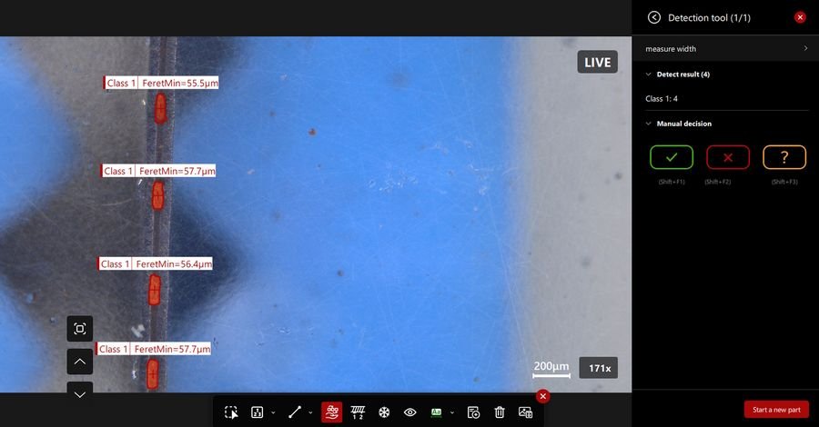
With AI-based visual inspection software, microscope users benefit from automated defect detection, classification, and measurement. It uses edge-learning models trained on image data. Unlike rule-based artificial intelligence, these systems learn patterns and adapt to complex or variable defects. With edge learning, a subset of machine learning, trained models run locally on the system where data is generated. It enables real-time inspection without cloud or internet dependence. Inspection workflows across production environments can be improved with the Aivaro AI Feature Detection software module. It reduces variability, improves consistency and efficiency and helps users make reliable quality decisions. A key requirement for defect detection during quality control is consistency. AI-based inspection supports this need by applying the same criteria to every sample. Once a model is validated, it can be reused across different systems and users, minimizing operator-dependent variation. Inspection results are stored with the image data, enabling traceability and documentation, which is particularly relevant in environments where results must be reviewed or audited. By standardizing both defect analysis and documentation, the Aivaro AI Feature Detection software module helps users achieve reproducible defect detection. For practical adoption, AI defect detection solutions must fit into existing inspection workflows. They should integrate readily into established quality control environments, without requiring major changes. For example, existing microscope solutions can still be used without changes to the SOPs, with the final decision remaining with users. The Aivaro AI Feature Detection software module can be implemented into workflows for inspection, quality control, failure analysis, and R&D, enabling users to achieve reliable defect detection. Solutions such as the Aivaro AI Feature Detection software module can be integrated into existing inspection workflows to support practical, scalable defect detection, enabling users to decide pass or fail rapidly and reliably.
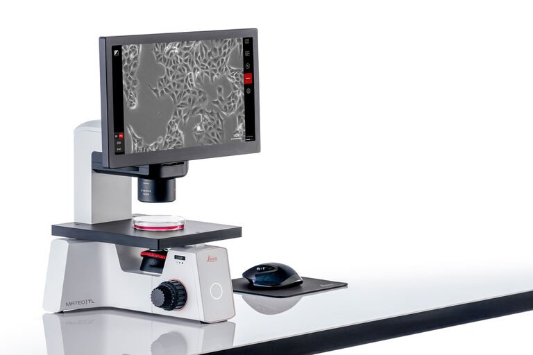
For researchers who need consistent experimental outcomes, Mateo TL enables all lab members to check and document cell growth status conveniently and comfortably. Measure confluency consistently, thereby increasing the confidence in the success of their downstream experiments. Comparison: Transmitted light - included; Fluorescence - not available; Confluency measurement - included; Cell counting - not available; Transfection efficiency - not available; Video output - not available. Mateo TL Digital Transmitted Light Microscope. Routine cell culture? Check! Empower your entire team to observe and take images of samples easily and intuitively. Simple to use, also for non-experts - All lab members, regardless of expertise, can set up contrasting methods correctly using the interactive assisted contrast setup feature - Observe and take images of samples easily with the intuitive and user-friendly software interface - Mateo TL eases the training burden on technicians, supervisors, and senior researchers Intuitive setup & use Mateo TL makes the process of cell culture checks effortless for every team member, with intuitive setup and use. It improves reproducibility for your experiments through objective confluency evaluation across individuals or experiments. Go from setup to first image in under 1 minute Benefit from a ready-to-use system which you can start working on right away. In addition, for cell culture work in regulated environments, once installed and qualified by our service team, you can start using Mateo TL immediately with consistent and reliable results from the start. You can add Installation Qualification (IQ) and Operational Qualification (OQ) to the service plan, which helps to document and verify that Mateo TL is installed and operating according to Leica’s specifications. One common criterion for consistent confluency measurements Mateo TL offers one common criterion for measuring confluency across the entire lab. The confluency algorithm measures the percentage of area covered by cells in the image. Mateo TL confluency module enables you to: - Eliminate subjective estimations - Achieve consistency in your confluency measurements between different users and experiments - Take the guesswork out of confluency evaluation - Use a common tool across lab experiments to consistently carry out confluency measurements and reduce variations - Improves experiment reproducibility - Minimizes human bias by using the confluency module at the point of decision https://www.leica-microsystems.com/fileadmin/ processed /d/5/csmMateo-Hood-nc8b21a6d29.jpg Smart functionalities Observe, take images of samples and export images quickly and conveniently using enhanced smart functionalities - Wirelessly transfer images directly to your smartphone or tablet with the quick share feature - Easily reproduce the imaging conditions of a previous reference image in the gallery using the repeat function - Free up more time from sample observation by eliminating the need to make frequent adjustments to the light and camera settings for each magnification with Mateo TL's built-in light memory function Enhance your team's comfort - Minimize fatigue by eliminating the need to use oculars - Mateo TL can be placed inside a laminar flow hood of compatible dimension for convenient cell culture work inside the hood, which also helps to minimize cell contamination
CATEGORY
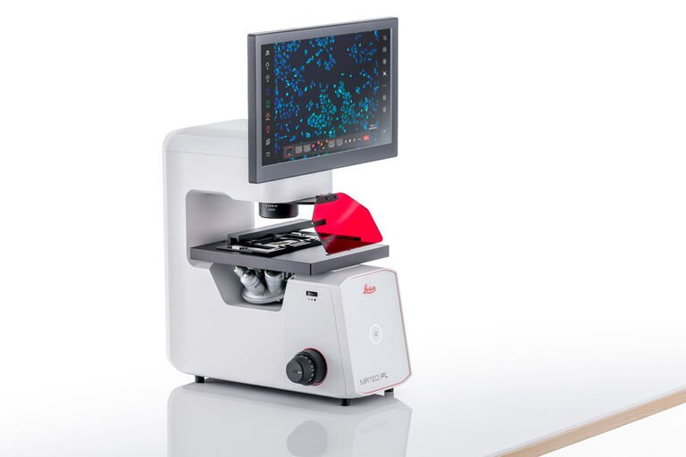
Boost your advanced cell culture research with Mateo FL, a digital inverted microscope with multi-modal fluorescence and transmitted light capabilities, automated analysis tools and secure data tracking. Reduce downtime and contamination risks using the integrated modalities and dual camera that minimize the need for sample transfer mid-experiment. Mateo FL Digital Fluorescence Microscope. Reliable, reproducible, advanced cell checks. Make your cell culture experiments more reliable and consistent with AI-powered algorithms and analysis. AI-powered workflows - Eliminate guesswork with AI-assisted confluency measurements. - Get an accurate, automated cell count that takes just 5 seconds per image with Mateo FL, saving you an average of 15 minutes of manual cell counting. - Optimize your transfection process with smart insights. Save time and effort with multi-modal imaging Mateo FL offers multi-modality fluorescence and transmitted light imaging in a single platform, saving time and providing flexibility in experimental design by optimizing each channel for specific fluorophores. See more with less fuss Enhance your visibility of unstained, transparent, translucent or low-contrast specimens with automated phase contrast. With a built-in dual camera system, it eliminates the need for physical camera changes and alignment, streamlining processes and improving efficiency. Everything you need in a single platform: multi-modal fluorescence and transmitted light imaging at a glance. Phase Contrast - Brightfield - Fluorescence (from left to right). Prevent sample contamination for consistent results Elevate your cell culture research outcomes by avoiding the inconvenience of contamination-related lab downtime. - Mateo FL can be used in biosafety level BSL 1 and BSL 2 laboratories as it is compatible with UV, ethanol and hydrogen peroxide decontamination methods. - Protect your sample from contamination and save valuable experiment time thanks to streamlined workflows and an integrated dual camera, eliminating the need for sample transfers in and out of the hood. Optimize your space and workflow with an integrated design The compact, integrated design of Mateo FL helps you to make the most of your bench space and make your workflow more efficient. Reduce contamination risks by at least 50% and double your workflow efficiency by capturing both monochrome and color images without needing to physically swap devices mid-experiment. Experience secure data tracking and seamless data transfer Mateo FL has built-in audit trails and user management functionalities that support FDA 21 CFR Part 11 compliance, making data management easier and more secure. - Effortlessly track and manage your samples with the bar code reader function. - Collaborate and share data more easily by seamlessly transferring your microscope data to any mobile device with QR codes. - Say goodbye to frequent file transfers or deletions - store up to 3 million images effortlessly with an ample 500 GB of built-in storage space. Comparison: Transmitted light - included; Fluorescence - included; Confluency measurement - included; Cell counting - included; Transfection efficiency - included; Video output - included.
CATEGORY

Digital microscopes are optical microscopes without eyepieces which display the image directly on a monitor. They enable suppliers and manufacturers to do investigation fast and share and communicate findings efficiently with each other, especially when current components are changed or new ones introduced. They also facilitate the easy documentation of results and creation of reports which can be referred to at a later time if the need arises. Digital microscopes are versatile and can be used for the investigation of diverse materials, from bigger, less sophisticated parts, such as tires and interior panels, to smaller, more complex, multi-component parts like PCBs (printed-circuit boards) and microelectronic chips. These attributes of a digital microscope allow an efficient workflow for investigation and documentation. Why are investigation and documentation efficient with a digital microscope? Here are some reasons: Go quickly from an overview to fine detail due to powerful lateral (XY) image stitching and a simple way to change magnification over the entire range (these videos on overview and mag change demonstrate how easy it is); Versatile illumination to see a part's diverse characteristics using the fully integrated ring and coaxial light; Save time with fast recall of imaging parameters thanks to the microscope encoding; Users need little training and experience to operate the microscope; View a part from multiple perspectives due to the fast microscope head tilting and sample stage rotation (to see how it is done, watch this video on tilting and rotation); Multiple users can easily operate the microscope for a variety of applications; Obtain comparable results no matter the microscope user thanks to the encoding and easy report generation. Table 1: Users can efficiently investigate and document auto parts with the DVM6 digital microscope thanks to a convenient way to: 1) change magnification rapidly over the full range, 2) tilt and rotate, 3) use the versatile, integrated illumination, 4) obtain reliable results, and 5) operate the microscope with little training. Fast Large Overview 2D Imaging (large to small field of view) X-Y Stitching; Quick & Easy Magnification Change 16:1 Zoom Optics Low, Mid, High Mag Objectives 12x to 2,350x; Change Viewing Perspective Rapidly Eucentric Tilting, +/-60 degree Sample Rotation, +/-180 degree; Fast 3D Models 3D Imaging Extended Depth of Field; Versatile Contrast Methods Integrated Ring & Coaxial Light Illumination; Easy Operation by Multiple Users Intuitive LAS X Software
CATEGORY
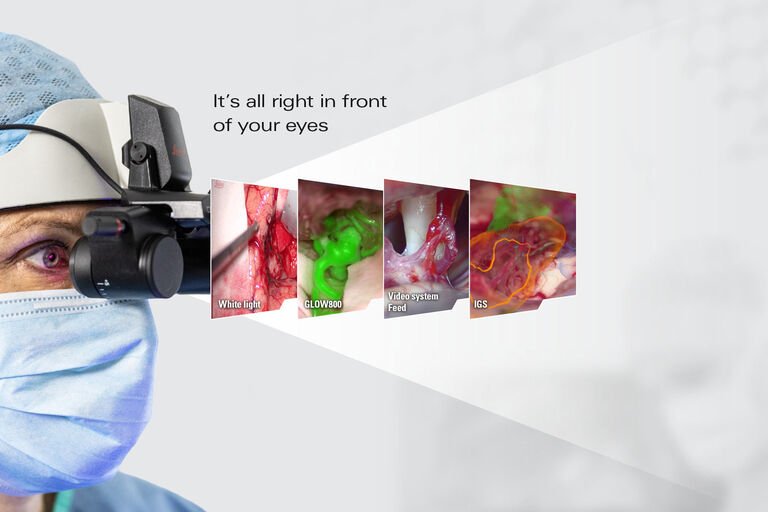
MyVeo for Neurosurgery, Spine surgery, PRS and ENT surgery Experience a smooth workflow during your procedures by not having to look at multiple screens for the information you need for clinical decision making. With MyVeo, you can access a broad spectrum of surgical information in real time in a high- resolution 3D view, at a click of a button on the microscope handle or via the foot pedal. This includes: - The white light image of your operating field - Augmented Reality applications - Image-guided surgery (IGS) data - Video system feeds from an endoscope Applications from external systems, such as IGS or information from compatible endoscopic video systems are only displayed in 2D resolution. OR team members can choose from four different image orientations to experience the surgery live with the MyVeo headset. They can either see from the same perspective as the main surgeon in high-resolution 3D or choose from these orientations: 90° rotated view in 2D, 180° rotated view in 3D, or 270° rotated view in 2D. Applications from external systems, such as IGS or information from compatible endoscopic video systems are only displayed in 2D, independent of the selected image orientation. Experience ergonomic comfort and ease of movement during long surgeries with the right view constantly in front of your eyes. With the MyVeo surgical visualization headset, you can: - Free yourself from the oculars or external monitors - Achieve a comfortable upright working posture - Connect instantly to surgery, with no waiting time - MyVeo is an interchangeable viewing option for the ARveo 8 microscope. In addition, MyVeo provides ease of movement with the freedom to easily flip up the lightweight near-eye screens of the headset when needed. MyVeo enables your team to visualize the entire surgery in 3D, at the same time as the surgeon. With fewer distractions and no crowding around monitors, a smooth surgical workflow is now possible. Up to three MyVeo users can experience real-time surgery, which is especially useful and powerful for teaching and learning. The MyVeo 3D surgical visualization headset is also designed to help you stay connected to your team with an unobstructed peripheral sight. You can still see the patient, your hands, the instruments, and your colleagues. MyVeo for Ophthalmology Experience a new dimension in 3D eye surgery with a single integrated view. MyVeo optimizes your workflow with fewer distractions - thanks to its immersive, high-resolution 3D view right before your eyes. Your view is complemented by EnFocus intraoperative real-time OCT imaging and phaco system information. With its large peripheral view, MyVeo keeps you visually connected to your patient, your instruments, and your team, without losing focus. Free yourself from the constraints of having to operate in a fixed position either via the microscope eyepieces or a 3D screen. Experience ergonomic comfort and freedom of movement, for cataract, retina and all ophthalmic surgeries, with the right view in front of your eyes. The MyVeo surgical headset supports a more natural posture, which is particularly beneficial to reduce neck strain. For ophthalmology, you can use MyVeo exclusively with the Proveo 8x 3D digital microscope to revolutionize the experience for your residents and impact the collaboration across your OR team. Using MyVeo headsets, your residents stay highly engaged with an undisturbed live view of the surgical field right in front of their eyes. It is possible for three headsets to be connected simultaneously.
CATEGORY
