By JEOL Ltd.
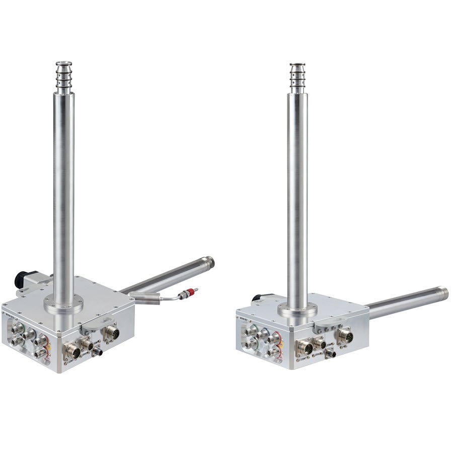
JEOL cryogenically cooled NMR probes are designed for maximum flexibility and uptime. Unlike conventional cryogenic probes that require lengthy warm-up cycles before removal or replacement—resulting in hours of downtime and unnecessary thermal stress—JEOL cryogenic probes can be removed from the magnet while still cold. This unique capability enables rapid switching between cryogenically cooled liquid-state probes, room-temperature liquids probes, and solid-state NMR probes, allowing laboratories to adapt quickly to changing experimental needs. Paired with the JEOL Probe Lifter, probe exchange becomes fast, safe, and effortless, helping researchers maximize instrument productivity and operational efficiency.
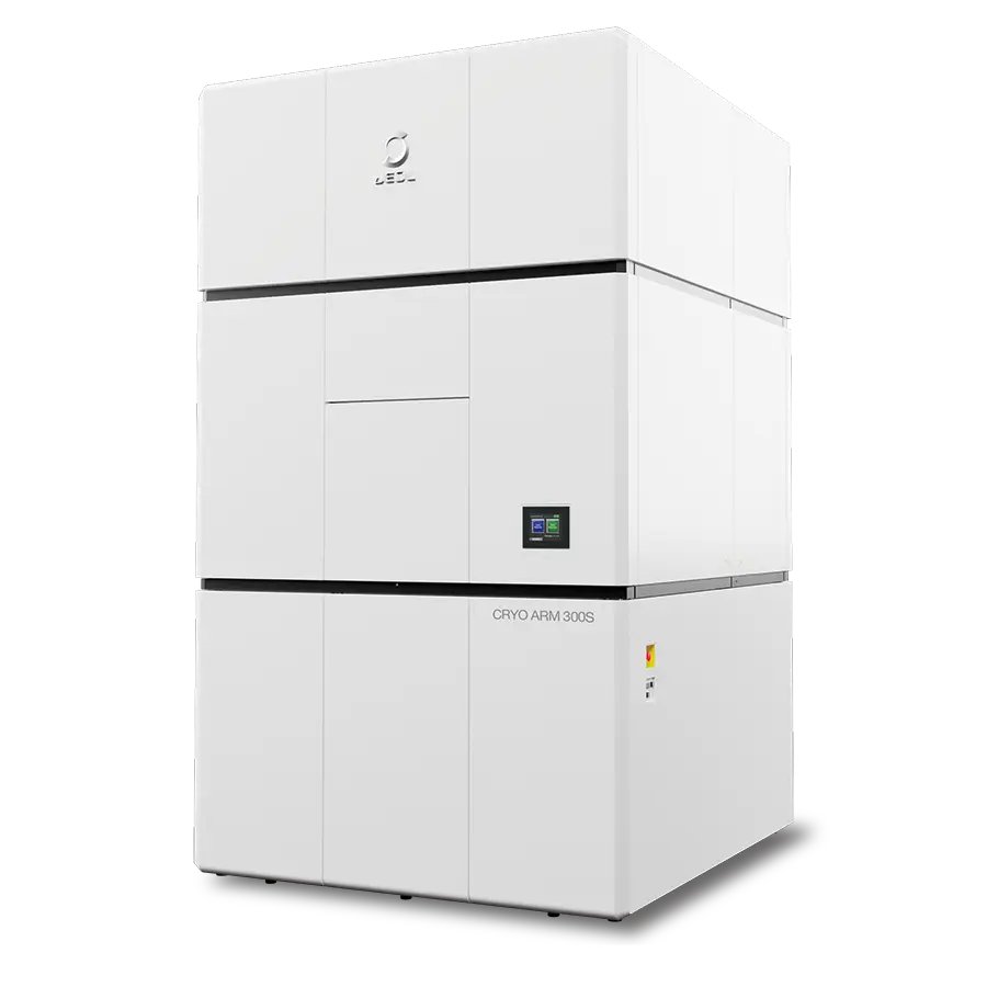
Field Emission Cryo-electron Microscope One cryo-electron microscope for single-particle analysis, cryo-electron tomography and 3DED/MicroED AI-driven automation and higher throughput make JEOL cryo-EM faster, more reproducible, and accessible to every operator. JEOL's latest 300 kV cold field-emission cryo-electron microscope that offers single-particle analysis (SPA), cryo-electron tomography (cryo-ET), 3DED/MicroED and beyond, all in one highly automated instrument. The CRYO ARM™ 300S is engineered to deliver high-resolution structural data with the stability, throughput and ease of use that today's structural-biology and drug-discovery laboratories require. CRYO ARM™ 300S × SmartScope Single particle analysis data acquisition supported by AI SmartScope is an open-source software that supports data acquisition in single particle analysis. Using AI, it automatically detects areas suitable for imaging and can automate the entire process up to data acquisition. In addition, with an intuitive UI that organizes the operation flow in a simple manner and with necessary configurations kept to a minimum, even beginners can achieve stable data acquisition regardless of the level of experience. CRYO ARM™ 300S × System Innovations High throughput, easy operation Smart sample transfer system CRYO SPECPORTER™-S The advanced CRYO SPECPORTER™-S separates the storage for sample preservation from the transfer magazines used for sample exchange. This permits selective handling of only the samples required during analysis, eliminating the need to load and unload all samples in the instrument during exchange. This supports stable and efficient operation, even in multiuser environments. Enhanced cartridge handling with higher transfer capacity The number of cartridges that can be loaded into a magazine at one time has expanded from four to six, reducing the frequency and time required for sample exchange, boosting throughput while maintaining stable sample storage environment. "Endless" multi-grid imaging capability A flexible sample transfer system allows selective, individual exchange of only the required samples. In multi-grid imaging, samples analyzed can be exchanged as needed while new samples are preloaded in advance, allowing uninterrupted overnight operation and maximizing data acquisition efficiency. Sample storage in a clean environment The storage is maintained under vacuum and at liquid nitrogen temperature, enabling sample storage for approximately one month in a clean, ice-accumulation-free state. This allows flexible sample management in accordance with analysis schedules. Beam designed for maximum information yield CRYO ARM™ 300S features fringe-free illumination, which prevents interference fringes during imaging. By making efficient use of the beam, data can be acquired with high throughput while minimizing damage to the sample. Improved flexibility of the electron beam Improvements to the illumination system, including the condenser lens (CL) and CL aperture, refine the flexibility and stability of the electron beam. Enhanced lens demagnification ratio and aperture positioning accuracy improve beam positioning at the specimen plane. Eight aperture sizes and seven optimized fringe-free beam conditions for each aperture size enable ideal illumination across a wide range of specimens. Longer consumable life for reduced maintenance Roots pumps are used for their extended maintenance intervals, reducing downtime and ensuring high system uptime. CRYO ARM™ 300S × CryoLameller Cartridge-based cryo-CLEM workflow delivers high efficiency and high reproducibility Integration with the CryoLameller (CRYO-FIB-SEM) enables a workflow that seamlessly connects sample preparation and analysis. From sample processing to data acquisition, the same cartridge holding the grid can be used throughout the workflow, allowing continuous operation while preserving positional information. With an integrated fluorescence microscope, intuitive and efficient area selection and positioning are achieved using correlative light and electron microscopy (CLEM). Specifications Main instrument Electron Gun: Cold field-emission electron gun Standard accelerating voltage: 300 kV Energy Filter: In-column Omega energy filter Options - Low accelerating voltage (Please make a separate inquiry about requests for other accelerating voltage except standard) - STEM unit - STEM detector (Bright-field/Dark-field) - Camera system (Direct electron detector and related equipment) - Smart Operation Software (AERO) - EDM (Electrostatic Dose Modulator) Specimen stage / Automated specimen exchange system / Specimen storage Specimen Stage Coolant: Liquid nitrogen, built-in automated liquid nitrogen filling system Cooling temperature: 100 K or less Specimen movements X, Y: Motor-driven (travel range: ±1 mm) Z: Motor-driven (travel range: ±0.2 mm) Tilt-X: Motor-driven (tilt range: ±70°) Specimen exchange system: Air-lock, built-in automated cryotransfer system Coolant: Liquid nitrogen, built-in automated liquid nitrogen filling system Cooling temperature: 105 K or less Specimen exchange cartridge: Up to 6 specimens can be exchanged at once. Specimen storage: Capacity of up to 12 specimens
CATEGORY
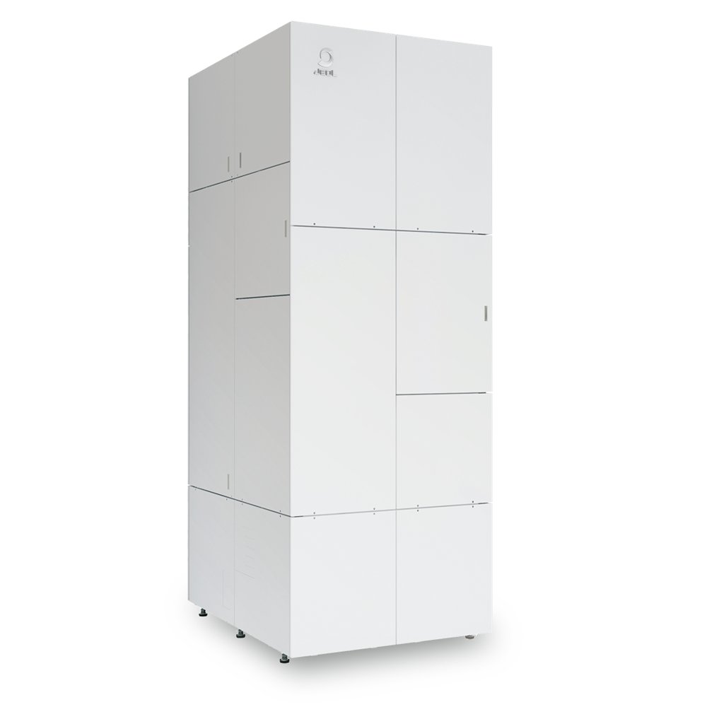
The JEM-Z200MF is an electron microscope with magnetic field-free objective lens which enables high resolution observation without applying strong magnetic field to specimens. Combined with a higher-order aberration corrector, atomic resolution imaging is possible. Key Features Magnetic field-free Objective Lens JEM-Z200MF objective lens consists of two lenses (FOL/BOL) located above and below the sample. The magnetic fields of these two lenses are canceled out at the sample plane, creating an objective lens with a field-free environment and a short focal length. The short focal length leads to low chromatic aberration and high overall stability. Combined with a higher-order aberration corrector, atomic resolution imaging is possible. A magnetic field in the Z-direction can be applied to the sample using built-in coils, located around the lens itself. Double Corrector The JEM-Z200MF is configured with Double corrector system on the illumination side as well as the imaging side, enabling both high-resolution STEM and TEM imaging. Illumination System The STEM corrector can be operated in two modes, one suitable for high-resolution observation, the other ideal for high sensitivity DPC investigations. Large convergence angle mode Ideal for high resolution observations. 200 kV, MHRP Convergence angle:20 mrad (semi-angle)with 40 μm CL aperture Small convergence angle mode Ideal for DPC STEM observations. 200 kV, MHRP Convergence angle: 1.0 mrad (semi-angle)with 10 μm CL aperture Imaging System With the JEM-Z200MF, it is now possible to switch between CV(:Conventional) Mode for dark-field/bright-field observation HR(:High-resolution) Mode by pressing a single button. Specifications Acceleration Voltages: 200 kV/80 kV Electron source: Cold-field Emission STEM Resolution - Bright-field: MHRP Configuration 0.092 nm; MWGP Configuration 0.136 nm STEM Resolution - Dark-field: MHRP Configuration 0.092 nm; MWGP Configuration 0.136 nm TEM Resolution - Lattice Resolution: MHRP Configuration 0.09 nm; MWGP Configuration 0.10 nm TEM Resolution - Information Limit: MHRP Configuration 0.14 nm; MWGP Configuration 0.16 nm Sample Tilt (X° / Y°) - Standard double-tilt holder: MHRP Configuration ±12° / ±16°; MWGP Configuration ±25° / ±27° Magnetic field around the sample: 0.3 mT or less Options: Energy-dispersive X-ray spectroscopy (EDS), Electron energy-loss spectroscopy (EELS), digital camera MHRP: Magnetic field-free High resolution Pole-piece MWGP: Magnetic field-free Wide Gap Pole-piece Applications & Gallery Visualizing electric/magnetic field distributions on an atomic scale Differential Phase Contrast imaging (DPC) Electric and magnetic fields can be visualized using the DPC-STEM method, which measures the small deflections of the electr on beam due to the fields inside the sample with segmented or pixelated detectors. Principle of DPC-STEM Observation of the magnetic field distribution (room temperature) of antiferromagnetic hematite at atomic resolution using DPC STEM. The symmetry of the hematite crystal was used to remove the information about the electric field and the magnetic distribution was visualized by averaging over all unit cells. Colors indicate the direction and strength of the magnetic field. Y. Kohno et. al, Nature 602, 234 (2022) Reduction of diffraction contrast effects in DPC-STEM Tilt-Scan system The JEM-Z200MF is equipped with a dedicated beam deflection system making it possible to change the incidence angle of the electron beam. Acquiring multiple DPC STEM images at different incident angles and then superimposing the individual images reduces the effect of diffraction contrast as shown below. (tilt-scan averaged DPC STEM, tDPC-STEM). Principle of tDPC-STEM Comparison of DPC TEM image of Nd 2 Fe 14 B with and without the use of the Tilt-Scan system. These images were observed along the axis of easy magnetization. Arrows in images indicate domain wall positions. Using the Tilt-Scan system diffraction contrast caused by precipitates is strongly reduced and the domain wall boundaries can be clearly observed. Gallery: High resolution STEM The combination of the magnetic field-free objective lens with a probe side Double corrector system enables atomic resolution STEM under magnetic field-free conditions. The large convergence angle mode enables STEM observation with a high spatial resolution on the order of 0.1 nm. Neodymium Iron Boron magnet (Nd 2 Fe 14 B) High-resolution HAADF-STEM image taken along the [001] direction and corresponding FFT pattern. The displayed orange circle corresponds to information transfer at 1Å. The STEM image was formed by summing 30 drift-compensated images. T. Seki et. al, Incommensurate grain-boundary atomic structure, Nature Communications 14, 7806 (2023), Fig. 1 Gallery: High resolution TEM in HR mode The HR mode enables TEM observations at higher magnification under magnetic field-free conditions. Combined with an image side spherical aberration corrector, atomic lattice resolved TEM images can be obtained. High-resolution TEM images of magnetite nanoparticles (Fe 3 O 4). By correcting the spherical aberration of the objective lens, the atomic arrangement of the magnetic particles can be studied. Gallery: Bright and Dark Field TEM image in CV mode Linking physical and magnetic structural features is made easy by JEM-Z200MF. In the CV mode, the diffraction plane aligns with the objective aperture plane, allowing for easy selection of diffraction features to generate bright field and dark field images of magnetic materials. Bright-field and dark-field images of dislocations in a pure iron sample. Dislocations were introduced by deforming the sample by 5% at liquid nitrogen temperature. Yellow circles indicate the positions of the objective aperture. Incidence Direction: Near <001 Data courtesy: Prof. Kazuto Arakawa, Shimane University Gallery: Lorentz-TEM / Fresnel method Using a TEM with a conventional objective lens, it is necessary to turn off the objective lens in order to observe magnetic domain structures. With JEM-Z200MF, magnetic domains are easily observed with the objective lens normally excited. Under focused image / defocus value -800 μm Observation of magnetic domains in a permalloy thin film (Fe 22 Ni 78) using the Fresnel method. Black and white arrows indicate the positions of domain walls. Fig. a Remanent state of the permalloy thin film Fig. b-d Using the magnetic field generating coils, which surround the objective lens, an external magnetic field is applied along the Z-axis. Sample courtesy: Dr. Takumi Sannomiya, Tokyo Institute of Technology
CATEGORY
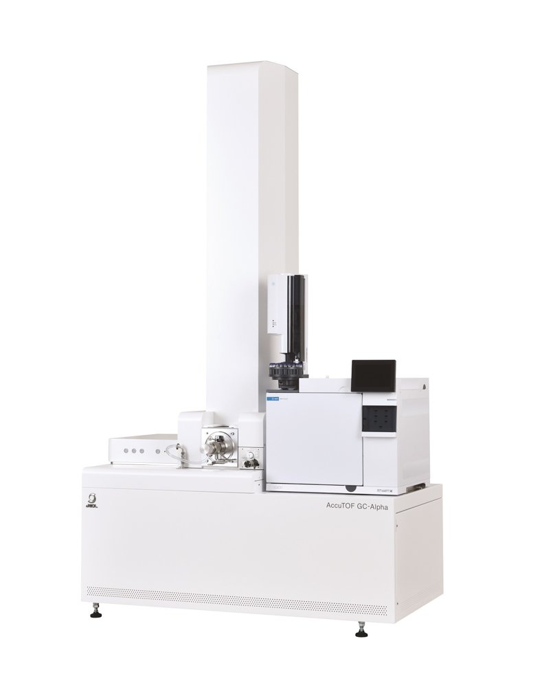
When Analytical Challenges Become More Complex Analytical laboratories are increasingly challenged to identify unknown compounds, analyze complex mixtures, and distinguish chemically similar materials. Many investigations require more than simply detecting compounds. Scientists often need to determine the identity of compounds and understand their chemical properties. For many samples, conventional electron ionization (EI) and mass spectral library searches alone may not provide sufficient information for compound identification. As a result, additional analytical information is often needed to fully interpret the results. Whether supporting research, quality control, failure analysis, environmental studies, or product development, laboratories need technologies that deliver deeper insight into increasingly complex samples. GC-Alpha: Identify Unknowns with Confidence GC-Alpha was designed to address these analytical challenges. By combining high-resolution accurate-mass GC-TOFMS, multiple ionization techniques, and intelligent software tools on a single platform, GC-Alpha provides complementary analytical information for confident chemical characterization and identification. Whether identifying unknown compounds, investigating degradation products, performing deformulation or failure analysis, or analyzing complex mixtures, GC-Alpha enables scientists to gain deeper molecular insight into their samples, helping answer analytical questions that would otherwise be difficult to resolve. Together, these complementary capabilities provide a powerful analytical approach to investigating complex chemical questions. What You Can Achieve with GC-Alpha Deeper Molecular Insight GC-Alpha provides multiple independent analytical data points, helping scientists better understand the chemical composition and characteristics of complex samples. Broader Structural Information By combining complementary ionization techniques, GC-Alpha provides molecular and structural information that would be difficult to obtain with a single ionization method. From Unknown Peaks to Identified Compounds Using msFineAnalysis AI, intelligent structure analysis software, GC-Alpha enables scientists to identify compounds, including peaks that conventional mass spectral library searches cannot identify. Flexibility Across Industries From biological/life science to plastics/polymer, energy, chemistry, food/plant, flavor/fragrances, environmental, semiconductor, petroleum and petrochemical, and academic laboratories, GC-Alpha offers the flexibility to address a wide range of analytical applications. The ultimate GC-MS system that pursues the ultimate in performance and functionality, the JMS-T2000GC AccuTOF™ GC-Alpha, has evolved into version “2.0” with the integration of a fully automated standard sample inlet and msFineAnalysis AI. It enables automatic structural analysis of unknown compounds from comprehensive two-dimensional gas chromatography (GCxGC) data, revolutionizing the conventional approach to qualitative analysis using GC-MS. Performance of AccuTOF™ GC-Alpha Aiming for high performance while keeping it simple, the AccuTOF™ GC-Alpha 2.0 uses two new Key Technologies. The AccuTOF™ GC-Alpha 2.0 is the 7th generation JEOL GC-TOFMS since the original AccuTOF™ GC. The AccuTOF™ GC-Alpha 2.0 employs orthogonal-acceleration time-of-flight mass spectrometer (oaTOFMS) with dual stage reflectron, an ideal ion optical system realizing both high ion transmission (= sensitivity) and ultra-high mass resolving power. A new workflow has been devised to identify unknown compounds by GC-MS. In this workflow, the data analysis is performed by integrating the data obtained by hard ionization (EI) and the data obtained by soft ionization (FI, PI, CI) in order to identify the analytes present in a sample. msFineAnalysis software streamlines the operation and adds a new function in which 2 similar samples can be directly compared for differential analysis. This new, enhanced version of msFineAnalysis allows the operator to easily accomplish qualitative analysis of unknown compounds as well as for compounds that are unregistered in the EI mass spectral libraries. High performance for both qualitative and quantitative analysis Four “High” specifications and two “Wide” specifications achieved simultaneously The JMS-T2000GC AccuTOF™ GC-Alpha 2.0 is a high-performance GC-MS system that simultaneously realizes high mass resolution, high mass accuracy, high sensitivity, high speed data acquisition, wide dynamic range, and wide mass range. The high mass resolution and high mass accuracy offers unprecedented qualitative analysis results. The high-speed data acquisition can be used for advanced GC-MS measurements such as comprehensive two-dimensional GC (GCxGC), while the wide dynamic range is useful for not only quantitative analysis but also for qualitative analysis of complex mixtures. The wide mass range is especially useful for direct MS measurements, and high sensitivity enables unprecedented information about trace components. The AccuTOF™ GC-Alpha 2.0 is truly a high-performance GC-MS system that removes the limitations for analysis. The standard EI ion source with ultra-high sensitivity is capable of trace quantitative analysis. Other ion sources are also sensitive enough for qualitative analysis and can be used in a wide range of applications. Eight sequential measurements of 100 fg of octafluoronaphthalene (OFN) were obtained by using the standard EI ion source.The instrument detection limit (IDL) is calculated based on the peak area and reproducibility of the extracted ion chromatogram for the molecular ion. An IDL of 18.7 fg was achieved for the system. OFN at concentrations ranging from 0.1 to 1,000 pg/uL (4 orders) were measured using the standard EI ion source and a high degree of linearity was confirmed. The wide dynamic range is useful not only for quantitative analysis but also for qualitative analysis of complex mixtures with different concentrations. GCxGC/EI TICC of diesel fuel For GCxGC and Fast GC analyses, the chromatographic peaks are very narrow, thus requiring a mass spectrometer that supports high-speed data acquisition. The AccuTOF™ GC-Alpha is a good match for these advanced chromatographic techniques because it can acquire data at up to 50 spectra per second. FD mass spectrum of polystyrene 5200 One of the features of time-of-flight mass spectrometers is their ability to measure a wide mass range. The upper limit of an ordinary GC-MS instrument is typically around m/z 1000, while the AccuTOF™ GC-Alpha can detect m/z 6000 and higher. This makes it possible to use direct probe MS methods such as field desorption (FD) to measure samples like oligomers. Mass separation of m/z 28 High mass resolving power is invaluable for qualitative analysis. Increasing the mass resolving power results in narrower mass spectral peaks, thus leading to the features below. Increased stability of the peak centroid = improved mass accuracy High mass accuracy makes it possible to determine the elemental compositions for the observed ions. Using the "drift compensation - multiple" mass calibration function, the average mass accuracy (absolute value) for 10 ions observed from methyl stearate was 0.05 mDa or 0.45 ppm. New Qualitative Analysis Capabilities offered by a Variety of Soft Ionizations AccuTOF™ GC-Alpha provides multiple ionization methods Soft ionization - a powerful tool for qualitative analysis EI, the most widely used ionization technique in GC-MS, is superior in sensitivity and reproducibility and is supported by a variety of library searchable databases. For any scientist using GC-MS in search of further information, what is ultimately important is molecular ion detection. EI, which uses a high ionization energy of 70 eV, generates numerous fragment ions, from which structural information is acquired. However, EI often fails to show strong molecular ion signals. Consequently, qualitative analysis results may be incorrect if they solely rely on database searches. Thus, it is critical in GC-MS analysis to enhance the accuracy of qualitative analysis by using various soft ionization techniques in addition to EI. Chemical ionization (CI), photoionization (PI), and field ionization (FI) are all optionally available on the AccuTOF™ GC-Alpha. With the accurate mass measurement capability for all ionization techniques, elemental composition of the analyte can be reliably determined. FI and FD are extremely soft ionization techniques that provide lower internal energy to the analytes than EI and even CI, thus producing clear molecular ions. As a result, FI and FD are ideal for molecular weight determination. FI (Field Ionization) - The sample is introduced to the ion source through GC or a standard sample inlet system. - Unlike CI, FI uses no reagent gas; no need to choose a reagent gas appropriate for the analyte. FD (Field Desorption) - The sample is applied onto the emitter and directly introduced into the system. - Suitable for analysis of thermally labile compounds. - Ideal for samples soluble in nonpolar solvents. - Analyzes powder samples dispersible in solvents. - Analyzes low- to mid-polar metal complexes. - Analyzes high molecular weight samples not supported in GC-MS, such as polymers. In FI and FD, ionization occurs by the removal of electrons from neutrals via the action of a high electric field. EI/FI/FD Combination Ion Source (optional) A single ion source that supports both EI (hard ionization) and FI/FD (soft ionization) techniques.Switching between EI and FI/FD is simple and quick. Features - No need to replace the ion source - No need to break the vacuum - No need for reagent gases The following analyses are possible by using this combination ion source with GC: - GC/EI for qualitative analysis through library search - GC/FI for molecular weight determination - Accurate mass measurement PI (Photoionization) ~ EI/PI Combination Ion Source (optional) PI is an ionization method that uses photons from a vacuum ultraviolet (VUV) lamp for ionization. The AccuTOF™ GC-Alpha has an optionally available combination ion source that offers both EI (hard ionization) and PI (soft ionization). This source makes it possible to switch between EI and PI by simply turning on/off the EI filament as well as the PI lamp. Features - No need to replace the ion source - No need to break the vacuum - No need for reagent gases Schematics of EI/PI Combination Ion Source PI is particularly useful for the analysis of aromatic compounds in complex mixtures. These compounds produce high intensity peaks with PI because they strongly absorb UV light. AccuTOF™ GC-Alpha with Direct MS Direct MS mode is useful for the analysis of high-boiling-point and high-mass compounds that are not amenable for GC. TOFMS with inherent wide mass range is well-matched with direct MS mode. Compounds that are measured by GC normally have molecular weights below 500 Da so it is not common to target compounds with molecular weights exceeding 1000 Da by using GC-MS. However, in direct MS mode (no GC is used), high-boiling-point, high-molecular-weight, and nonvolatile compounds can be targeted for measurement, as the sample is directly introduced into the ion source. The mass range of the AccuTOF™ GC-Alpha is m/z 6,000 or higher. Since compounds can be detected over a wider mass range than conventional GC-MS, the system is well suited for measurements in direct MS mode. DEP (Direct Exposure Probe) - Sample dissolved or dispersed in solvent is applied to the filament at the tip. - Suitable for high boiling point and/or thermally labile compounds - Compatible with EI and CI DIP (Direct Insertion Probe) - Solid sample can be introduced in the glass sample tube. - Suitable for high boiling point and/or insoluble compounds - Compatible with EI and CI FDP (Field Desorption Probe) - Sample dissolved or dispersed in solvent is applied to the carbon emitter at the tip. - Suitable for high boiling point, high molecular weight, and/or thermally labile compounds - Suitable for low- to mid-polar metal complexes - Used for FD soft ionization Applying the sample to the emitter In the FD method, the sample is dissolved or dispersed with a solvent and then applied onto the emitter to be introduced into the ion source. The sample does not need to be completely dissolved in the solvent. In this case, it is possible to analyze only the additives in the solvent. Since it is a direct sample injection method without using a column, high boiling point (high mass) components can be measured. Since it is soft ionization, molecular ions can be detected.A clear difference peak was detected at m/z 943.4797 from the flame-retardant PP. It was estimated to be a brominated flame retardant by isotope pattern analysis and composition estimation. Key Features High Sensitivity - High Mass Resolving Power: 30,000 - High Mass Accuracy: 1 ppm (based on our standard testing) - Instrument Detection Limit: IDL=18.7 fg - Wide Dynamic Range: 4 orders - High Speed Data Acquisition: 50 Hz - Wide Mass Range: ~m/z 6,000 Key Technologies New high-performance hardware The JMS-T2000GC AccuTOF™ GC-Alpha 2.0 is the 7th generation JEOL GC-TOFMS and has an improved ion optics system to achieve ultra-high resolution. The AccuTOF™ GC-Alpha is an orthogonal-acceleration time-of-flight mass spectrometer (oaTOFMS) with dual stage reflectron. It employs an ideal ion optical system realizing both high ion transmission (=sensitivity) and ultra-high resolution. Mass resolution and mass accuracy – Crucial for qualitative analysis The Resolving power of a time-of-flight mass spectrometer is calculated according to the formula above. With this equation in mind, the new JMS-T2000GC AccuTOF™ GC-Alpha hardware was designed to achieve ultra-high resolution using the following principles: - Make t longer: increase the flight distance 4 m - Make Δ t smaller: new ion optical system using dual stage reflectron - Make Δ t smaller: ion transfer system that can accommodate ions with a wide range of kinetic energy With the newly developed hardware, the AccuTOF™ GC-Alpha features 6 times higher resolution than the first generation AccuTOF™ GC and has a mass accuracy of ≤ 1 ppm. Acquiring unrivaled high-quality data, the AccuTOF™ GC-Alpha is the ultimate solution for GC-MS qualitative analysis.
CATEGORY
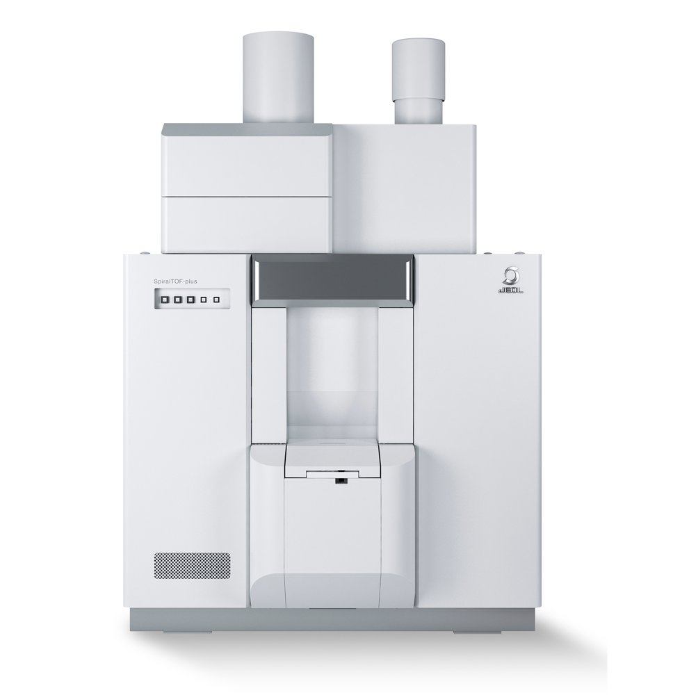
What sets NewSpiralTOF™ apart is its ability to deliver clean, high-sensitivity measurements in the low-mass region, where conventional MALDI reflectron TOFMS systems often suffer from chemical noise caused by the post-source decay (PSD) fragments of matrix cluster ions. Because the SpiralTOF ion optics are built from sector electric fields, low-kinetic-energy ions generated by PSD are effectively excluded before they reach the detector. The result is a mass spectrum with virtually no chemical noise in the low-mass region, enabling highly sensitive and accurate mass measurements of small molecules. Key Features Resolution, Accuracy, and Sensitivity All in One Instrument Feature 1: Ultra-high mass-resolution, ultra-high mass-accuracy Built on JEOL's proprietary SpiralTOF ion optics, derived from the MULTUM II ion optics system developed at Osaka University, the NewSpiralTOF™ achieves an effective flight path of 17 meters in a compact instrument design. This advanced ion optical configuration combines high ion transmission with performance levels that are exceptional for MALDI-TOFMS: mass resolution of 75,000 and mass-accuracy of 1 ppm using internal calibration. Feature 2: High quality data for everyone A longer flight path does more than improve performance under ideal conditions. It also enhances day-to-day practicality by reducing the influence of specimen-surface irregularities, such as matrix crystal morphology on a target plate or surface variation in mass imaging specimens. This makes it easier for users to obtain high-quality, reproducible data under real-world operating conditions. Feature 3: High sensitivity despite long flight path Even with a long flight path, NewSpiralTOF™ maintains excellent sensitivity. Ion packets of the same mass are refocused at each cycle of the spiral trajectory, allowing the system to achieve high mass-resolution and high mass-accuracy without compromising high sensitivity. Feature 4: Analysis of small molecules realized with very low chemical noise Because the SpiralTOF ion optical system, which consists of electric sectors, also acts as an energy filter, fragment ions generated by PSD are prevented from passing through the analyzer. This is the key to its remarkably low chemical noise in the low-mass region and its superior performance for small-molecule analysis. NewSpiralTOF™ High-Speed MS Imaging × High Mass-Resolution In MALDI mass spectrometry imaging (MALDI-MSI), molecules are visualized directly within a specimen by coating the specimen surface with matrix, scanning it with a laser, and acquiring a mass spectrum at each position. This makes it possible to see where specific molecules are located and how much of them is present across a tissue section or other specimen surface. High Mass-Resolution for Accurate MS Imaging Thanks to its 17 m flight path, the NewSpiralTOF™ delivers high mass-resolution even when analyzing biological tissue sections with nonuniform surface conditions. In measurements of approximately half of a mouse brain over a region of about 5 × 7 mm, the system achieved a mass resolution of approximately 40,000 in the average mass spectrum. This high resolving power enabled the separation of isobaric lipid species such as phosphatidylcholine (PC), phosphatidylethanolamine (PE), and galactosylceramide (GalCer), making it possible to obtain the correct spatial distribution for each molecule. AI-Enhanced Image Quality for MS Imaging JEOL has a long history of innovation in image processing, driven by its leadership in electron microscopy. Building on this expertise, JEOL adapted its AI-based image enhancement technology LIVE-AI (Live Image Visual Enhancer-AI), originally developed for SEM, to MS imaging data processing and implemented it as the FINE-AI Filter. The result is a substantial improvement in the clarity and interpretability of mass images. Automatic Extraction of Important Features With the high mass-resolution of the NewSpiralTOF™, more than 100 lipid species were detected in mouse brain sections. However, interpreting the distribution of each lipid individually is not practical. Meaningful insight requires statistical tools that can identify groups of components that vary together and summarize complex datasets efficiently. To address this, vertex component analysis (VCA), a method that models all spectra in a mass imaging dataset as mixtures of a small number of "vertex components" is implemented. Compared with methods such as principal component analysis, VCA provides results that are easier to interpret in a shorter time. Because VCA is sensitive to noise, applying it after denoising with the FINE-AI Filter makes the analysis far more effective. In the example shown, this approach revealed three key vertex components characterizing lipids in mouse brain tissue. Features and usages of TOF-TOF option and linear TOF option The TOF-TOF option extends the analytical power of the NewSpiralTOF™ by using SpiralTOF ion optics in the first MS stage, enabling high precursor-ion selectivity and accurate selection of the monoisotopic precursor ions. Combined with high energy collision-induced dissociation (HE-CID), the system generates product-ion spectra rich in structural information. JEOL's proprietary offset parabolic reflectron further strengthens TOF-TOF performance by allowing simultaneous acquisition of product-ion information from m/z 5 up to the precursor ion. This provides comprehensive fragment information and supports highly reliable structural analysis. A representative example is the analysis of reserpine and its photodegradation product. Because reserpine is photolabile, its solution degrades under ordinary room light. The degradation product differs by 2 Da, suggesting the loss of two hydrogen atoms. Thanks to the high precursor selectivity of the TOF-TOF option, the NewSpiralTOF™ successfully acquired separate product-ion spectra for both reserpine and its degradation product directly from a mixture, enabling structural characterization of each species. With the Linear TOF option, ions travel directly from the ion source to the detector. If post-source decay occurs during flight, the resulting fragment ions and neutral species continue at the same velocity as the precursor and are detected as the same signal. This makes Linear mode particularly effective for the high-sensitivity analysis of high-mass compounds that are prone to PSD. Used together, Spiral mode and Linear mode broaden the range of analytes that can be measured. The application examples are the confirmation of the molecular weight distribution of polymers above 100 kDa and the measurement of the molecular weight of immunoglobulin G (IgG), a glycoprotein of approximately 150 kDa. Stable Performance with a Contamination-Resistant Ion Source In MALDI ion sources, much of the desorbed matrix leaves the target as neutral particles, and some of this material adheres to the ion extraction electrode. In conventional MALDI-TOFMS systems, contamination can accumulate unevenly on the aperture lens, leading to uneven charging and deflection of ion trajectories. This degrades both sensitivity and mass resolution, making frequent cleaning necessary. To overcome this issue, the NewSpiralTOF™ uses a fine, high open-area ( 82%) ion extraction grid. Because less contamination accumulates overall, and because the ion-optical properties of the grid reduce the impact of any charge that does build up, ion trajectories remain stable even over long-term use. The result is maintenance-free operation with sustained high sensitivity and high mass-resolution. Analysis Examples NewSpiralTOF™ Life Science Applications Confident Protein Identification by Peptide Mass Fingerprinting (MSTips No.166) Peptide mass fingerprinting remains one of the most fundamental tools for protein identification. With its combination of high mass-resolution, high mass-accuracy, and high sensitivity, the NewSpiralTOF™ enables highly reliable protein identification with fewer false positives. High-Resolution Protein Measurement (MSTips No. 297) The system's high resolving power and sensitivity make it possible to measure intact proteins such as ubiquitin at high resolution, including clear detection of the protonated molecular ion. Rapid Sequence Confirmation by In-Source Decay (ISD) (MSTips No. 229) For rapid sequence confirmation of recombinant proteins, the in-source decay (ISD) method is highly effective. When used with MASCOT Server (including the Top Down option), the NewSpiralTOF™ can support sequence verification, as demonstrated for horse myoglobin. Structural Analysis of Phospholipids in Egg Yolk (MSTips No. 185) The TOF-TOF option and HE-CID capabilities of the NewSpiralTOF™ also support structural analysis of complex lipid mixtures, including phospholipids extracted from egg yolk. High precursor selectivity makes it possible to isolate the correct ion and obtain informative fragmentation patterns for structural characterization. Structural Analysis of a Derivatized N-linked Glycan (MSTips No. 519) In glycan analysis, labeling the reducing end with a fluorescent tag is common for HPLC workflows, and many such labels also improve ionization efficiency in MALDI and ESI. The structural analysis of glycans labeled with 2-aminobenzamide (2-AB) using the NewSpiralTOF™, including the TOF-TOF option, is shown below. Confirmation of a Synthesized Oligonucleotide (MSTips No. 364) The instrument's high mass-resolution and mass-accuracy also make it possible to confirm the elemental composition of synthetic oligonucleotides, expanding its usefulness into nucleic acid analysis. NewSpiralTOF™ Chemistry and Materials Applications End-Group Analysis of High-Molecular-Weight Polymers up to 100 kDa (MSTips No. 527) With a wide mass range of m/z 4 -120,000 in Spiral mode, combined with high resolution and high mass-accuracy, the NewSpiralTOF™ enables end-group analysis of polymers with molecular weights exceeding 100,000, including polycarbonate. Accurate Mass Measurement of Covalent Supramolecule of 9 kDa (MSTips No. 369) The system also supports accurate mass measurement of covalent supramolecules above 9,000 Da, with errors reported at below 0.1 ppm. This level of performance contributed to work published in the Journal of the American Chemical Society (JACS) .
CATEGORY
