
By NanoFCM
Implementing strategies for single particle quantification and cargo detection, NanoFCM provides a versatile and powerful platform with the NanoAnalyzer, a flow cytometry based system for multi-parameter nanoparticle analyses. Precise measurement of elastically scattered light from single nanoparticles allows for high-resolution size distribution, comparable to cryo-electron microscopy, with fast acquisition times of 1-2 minutes also yielding particle concentration data. Fluorescent labelling technologies can be implemented to yield concurrent detection of important cargos such as proteins, nucleic acids and lipids. Global implementation at some of the top research institutions and high-tech enterprises continue to demonstrate the benefits of nano-flow cytometry for particle characterization. NanoFCM's NanoAnalyzer instrument has become established as a powerful tool for multiparameter characterization of natural and synthetic nanoparticles at the single-particle level.
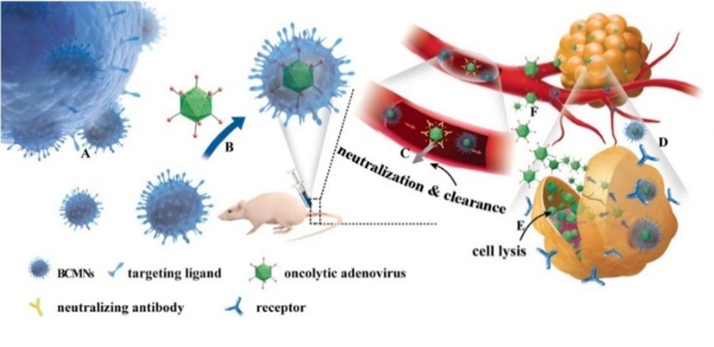
Each method provides separate important data about a lentiviral sample. Nano-flow cytometry can be employed to describe all these lentiviral characteristics simultaneously at a single particle level. Additionally, the use of the Flow NanoAnalyzer on lentiviral products identifies both mature lentiviruses and contaminant unincorporated components. Multiple components coexist in lentiviral samples, such as mature virions, partially assembled virions, free nucleic acids, etc. Analysis by nano-flow cytometry can give a thorough description of the active population of lentivirus presenting VSV-G for cellular recognition and infectivity, and internal nucleic acid cargo for activity. This data is highly quantitative, achieved rapidly, and simultaneously measures the other immature particles to determine product purity. Figure 1. Analyze different compositions of lentivirus particles on the Flow NanoAnalyzer In Figure 1, the P1 population represents the complete lentivirus, double-positive for labeled nucleic acid and VSV-G proteins, making up ~45% of the particle product, with additional size distribution and concentration data provided in a single 2-3 minute run time. The major particle contaminant is the VSV-G-negative, genome-positive population. Despite the potential for these VSV-G-negative particles to be insufficient for gene integration, they could skew qPCR measurements and particle counting methods. This study demonstrates that exosome-encapsulated oncolytic viruses exhibit antiviral immunity, and the targeting of tumor cells can be enhanced by labeling tumor-specific markers on the surface of exosomes. Releasing the oncolytic adenovirus into the exosomes is a key step in the experiment. The encapsulation efficiency and effectiveness of oncolytic adenovirus directly impacts the subsequent treatment outcomes. The researchers used fluorescent dye CFSE and nucleic acid dye SYTO™ 62 to label exosomes and OA, respectively. The Flow NanoAnalyzer was employed to evaluate the encapsulation efficiency of oncolytic adenovirus. Figure 1. Encapsulation efficiency of oncolytic viruses. Based on nucleic acid staining, the Flow NanoAnalyzer allows the determination of the encapsulation efficiency of oncolytic adenoviruses, which was 59.9% in this case. A high-throughput single-particle method to enumerate intact viral particles using the Flow NanoAnalyzer was reported. Using the bacteriophage T7 as a model system, intact virions were completely discriminated from empty capsids and naked viral genomes. Successful measurement of the physical virus titer and purity was demonstrated for recombinant adenoviruses, which could be used for gene delivery, therapeutic products derived from phage cocktails, and infected cell supernatants for veterinary vaccine production. Figure 1. Characterization of virus-based vaccines.
CATEGORY

Based on the superior sensitivity for single fluorophores, antibodies and mRNAs and equipped with automated system, Single-Molecule NanoAnalyzer brings the unprecedented fluorescent quantification into nanoparticle analysis. Single-Molecule Sensitivity FL signals of single fluorophores, antibodies, and mRNAs can be easily distinguished from background. Fluorescent Quantification Quantification of the copy number for carried nucleic acids and specific proteins. Automated System Automated cleaning and sampling system. Data Management The updated data management system (FDA 21 CFR Part 11 compliant) ensures data authenticity. Single PE Detection The scattering was as low as 24 nm and the fluorescence as low as single PE detection. Single Antibody Characterization The fluorescent signals of Alexa Fluor 647 conjugated Antibodies presented single and uniform signal during individually by nano-flow cytometry, which means the NanoFCM is able to analysis single antibody. Single mRNA Characterization The fluorescent signals of mRNA which stained with nucleic acid dye presented single and uniform signal during individually by nano-flow cytometry, which means the NanoFCM is able to analysis single nucleic acid. Superior Sensitivity in Single Molecule Analysis FL singnals of single florophores, antibodies, and mRNAs can be easily distinguished from background. Fluorescent Qutification Equipped with fluorescent quantitative nanobeads and one-click algorithm, Auto20 effortlessly achieves precise calculation of specific proteins and nucleic acid copy numbers in nanoparticles of interest. It brings forth novel solutions for targeted drug development and quality control. Automated System The automated sample loading and cleaning system brings unprecedented convenience to your experiments. Data Management The updated data management system (FDA 21 CFR Part 11 compliant) ensures data authenticity. Extracellular Vesicles Optimum configuration of multi-laser and multi-fluorescent channels by the Auto 20 can achieve the multiparameter analysis of the three tetraspanins (CD9, CD63 and CD81) for single EVs. mRNA LNPs Determination of the copy number of the carried mRNAs and the specific ligands for nanomedicines by high-resolution fluorescent quantification. Lentiviruses For the first time, simultaneous analysis of three key components (P24, VSV-G and mRNA) of single lentiviruses was achieved.
CATEGORY
