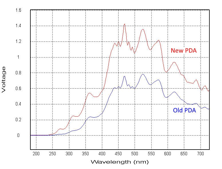
Increase detection sensitivity and efficiency Direct mounting of a new photodiode array detector to the cell block of the sample handling unit eliminates the need for a fiber-optic link between the cell and detector and improves overall light efficiency and sensitivity: - Greatly improved signal:noise in kinetic traces - Better absorbance signal accuracy in UV range - Improved spectral resolution with less noise - Faster PDA integration speeds (minimum 0.68ms) - Increased number of spectra per stopped-flow drive–16000 - Further improvements using deuterium lamp (optional). The figure shows a comparison of xenon lamp voltage output under identical conditions when using the old fiber optic and new direct-mount PDA accessories. Increased light throughput of the direct mount PDA allows use of a much lower gain setting, significantly reducing noise levels. Data shows the comparison at 350nm. “The direct mount PDA upgrade from Applied Photophysics allowed me to upgrade my existing PDA to the latest specification. The upgraded PDA is really simple to fit to the instrument, has given me a ten-fold increase in sensitivity and has greatly improved the signal to noise ratio of my enzyme measurements.” -Dr Andy Holt, University of Alberta. Compatible with all diode array models supplied to SX systems dating back to 1990. The existing Zeiss spectrometer unit from the original housing is transferred to the updated unit containing the latest control electronics and coupling optics. Some older models may require upgrading to Pro-Data Windows based software.
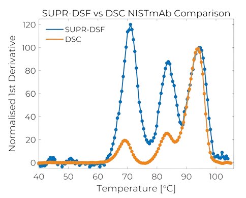
Next Level Differential Scanning Fluorimetry The SUPR-DSF by Protein Stable delivers next level differential scanning fluorimetry by simplifying sample preparation and analysis of protein stability into a single microplate. Utilising precision optical components to deliver the highest sensitivity, intrinsic fluorescence of proteins and full spectrum detection, both thermal ramping and isothermal chemical denaturation experiments can be easily run in 384-well microplates. Ensure reliability with our five-year warranty, providing extended protection and peace of mind. This additional coverage ensures long-term support, safeguarding your investment. Please note that for the warranty to be valid, you need an active support plan. Technology - High throughput thermal ramping stability screening - Intrinsic fluorescence - broad compatibility with biological buffers - Fast, 384-well plates scanned at 1 o C per minute - Wide dynamic range of sample concentrations - Very low protein requirement and volumes needed Applications - Formulation and buffer optimisation - Protein characterisation - Stability profiling - Similarity assessment - Accelerated stress and forced degradation studies - Binding-induced conformational changes analysis - Post-translational modification assessment Next generation differential scanning flourimetry Ease of use is at its core - The SUPR-DSF was designed specifically to deliver great protein stability screening whilst simplifying processes, thus reducing operator time in preparing samples, reducing the amount and cost of consumables required, and reducing the risk of errors in extra preparation steps. - By developing a system that can read directly in-plate, the shift in intrinsic fluorescence of protein samples as they unfold, Protein Stable has eliminated several steps seen in other techniques. - Thus sample preparation starts and stops in a single 384-well plate and once loaded onto the thermal block that moves out of the drawer at the front of the instrument, it is simply a matter of loading up a method and starting a run in the graphical SUPR-Suite software. Once complete, the analysis and export of the data are a few clicks away. Data says it all - Protein stability screens can reveal significant differences between samples’ melting temperatures, however the SUPR-DSF can be used to spot much smaller differences and more. - Using a spectrometer to detect full emission of the intrinsic tryptophan and tyrosine residues far greater detail can be elucidated. - Analysis options include several methods using either more traditional ratio methods of two wavelengths or more comprehensive methods using the spectrum. More data facilitates better signal to noise and more detail. On board automatic analysis breezes through 384 samples and should you wish to use external programs as preference, exporting data is straightforward. Efficiency for you - Preparing samples and subsequently analysing them in the same microplate certainly reduces the number of steps in the stability screening method, thus reducing operator interaction and risk of error, however the real efficiencies are realised in running costs. - All you need to run the SUPR-DSF is a standard black 384-well PCR plate and a plate seal. No extra vessels to load the samples into, no cartridges or structures to hold them in place. The extras can be significantly expensive. - Additionally, using a PCR plate allows sample volumes to be as little as 10 μL and with protein concentrations also being low, savings also extend to the sample you need to prepare.
CATEGORY
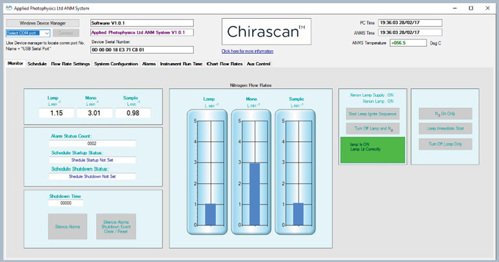
The laboratory standard circular dichroism instrument Suitable for studying chiral molecules of all types and sizes, this Chirascan CD spectrometer has featured in thousands of publications since its introduction in 2005. Often used to analyze biomolecules, Chirascan data contributes to a deeper understanding of biomolecular characteristics, mechanisms and interactions. More than α-helix and β sheet - determine structural and thermodynamic properties - Gain insight and detect changes in secondary and tertiary structure - Determine response to thermal or chemical change - Study folding and unfolding mechanisms - Expand capabilities with dedicated Chirascan accessories - Ensure reliability with our five-year warranty Features and accessories Chirascan systems are supplied with the features and accessories required for acquisition of high quality data from 'day one'. Molecular sieve, activated charcoal filter - Removes common gas impurities Active nitrogen management system - Regulates purge gas consumption - Software-controlled Air-cooled xenon lamp - Software-controlled - Up-time recorded Monochromator - Produces horizontally, linearly polarized monochromatic light - Dual polarizing prisms maximize light throughput Photoelastic modulator - Converts horizontally polarized light to circularly polarized light. Alternates between left- and right-handed circular polarized light Temperature-controlled sample chamber - Consistent analytical conditions - Continuous temperature ramps (single sample mode) Water circulator - Dissipates heat from sample chamber and sample storage Peltiers Cuvettes and holders - Selected for far- and near-UV CD analysis of biomolecules (single sample mode) Photomultiplier Detector Control and analysis software - Easily defined run parameters and store routine protocols - Saves time with scheduled start-up/shutdown of lamp and N2 supply - Fail-safe lamp switch-off if N2 flow drops - Ensures O2-free conditions with N2 purge - Recognizes flow cell to select optimal run/wash/dry protocol
CATEGORY
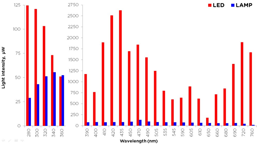
Increase sensitivity of fluorescence measurements The SX LED Light Source Upgrade provides a significant improvement in performance of stopped-flow fluorescence measurements. The higher intensity and stability of the LEDs compared to the existing xenon light source increases signal:noise and detection sensitivity limits. - Lower noise levels and increased sensitivity for all fluorescence modes - Simple to install and alignment-free - Long lifetimes (typically 5000-50000 hours) - Complement or replace older xenon light sources - Compatible with all SX and Chirascan stopped-flow models. Comparison of LEDs and xenon light source intensity The LED Light Source comprises a power supply and one or more of a selection of wavelength-specific LEDs covering the UV and visible region (280-760nm). The LED connects directly to the cell block of the sample handling unit in place of the fiber-optic light guide from the xenon light source and monochromator.
CATEGORY
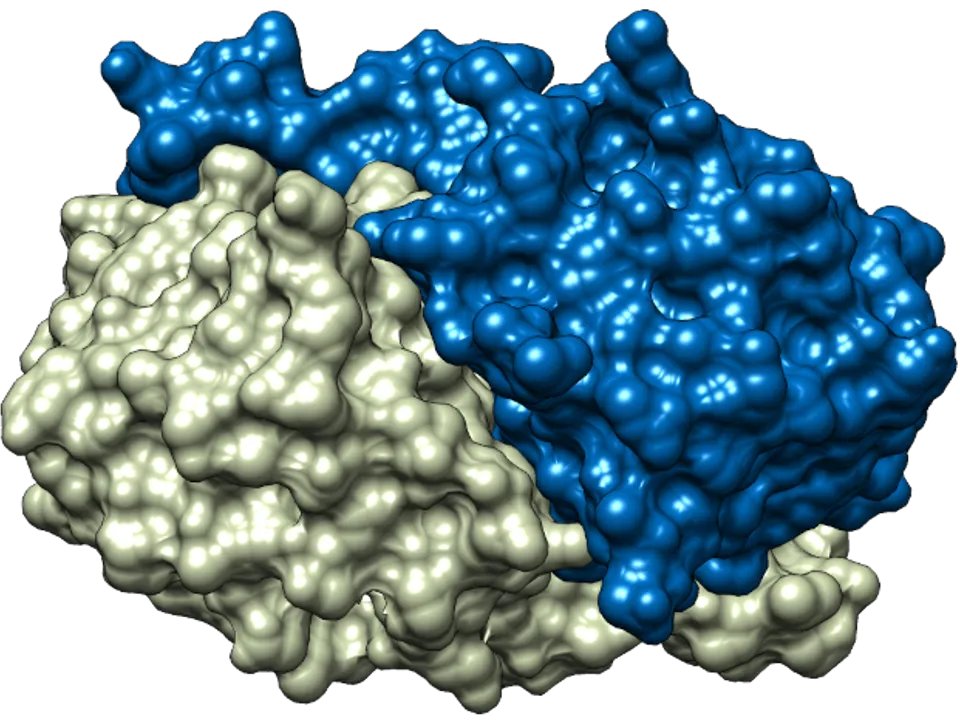
Stopped-flow experiments using Applied Photophysics SX20 and Chirascan V100 SF.3 systems allow monitoring of different spectral properties including fluorescence, absorbance, and even far-and near-UV CD.With these tools. This study outlines the transition between fully folded and locally unfolded states of Tsa1, in its sulfenic acid form CP-SOH, is not in rapid equilibrium as previously hypothesised, but is almost irreversible (kFF~0s-1)and in direct competition with the hyperoxidation step.
CATEGORY
