By Nanosurf AG
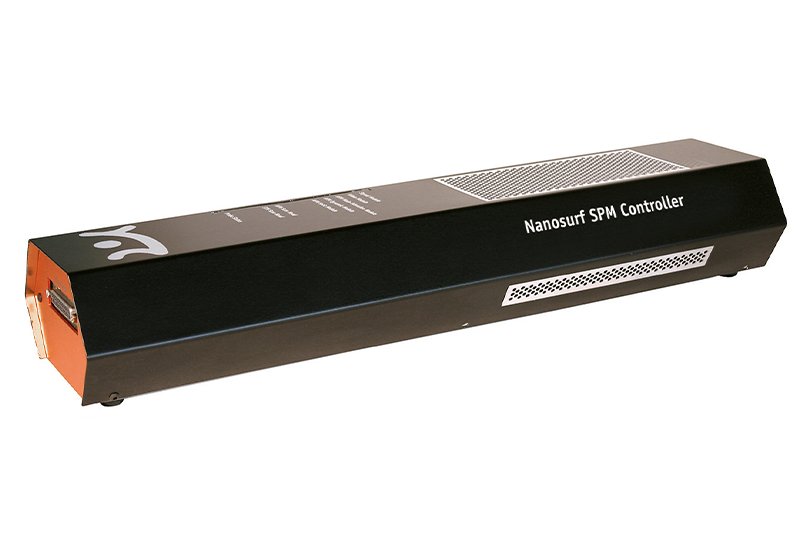
Download the control software for products with the SPM S Controller.
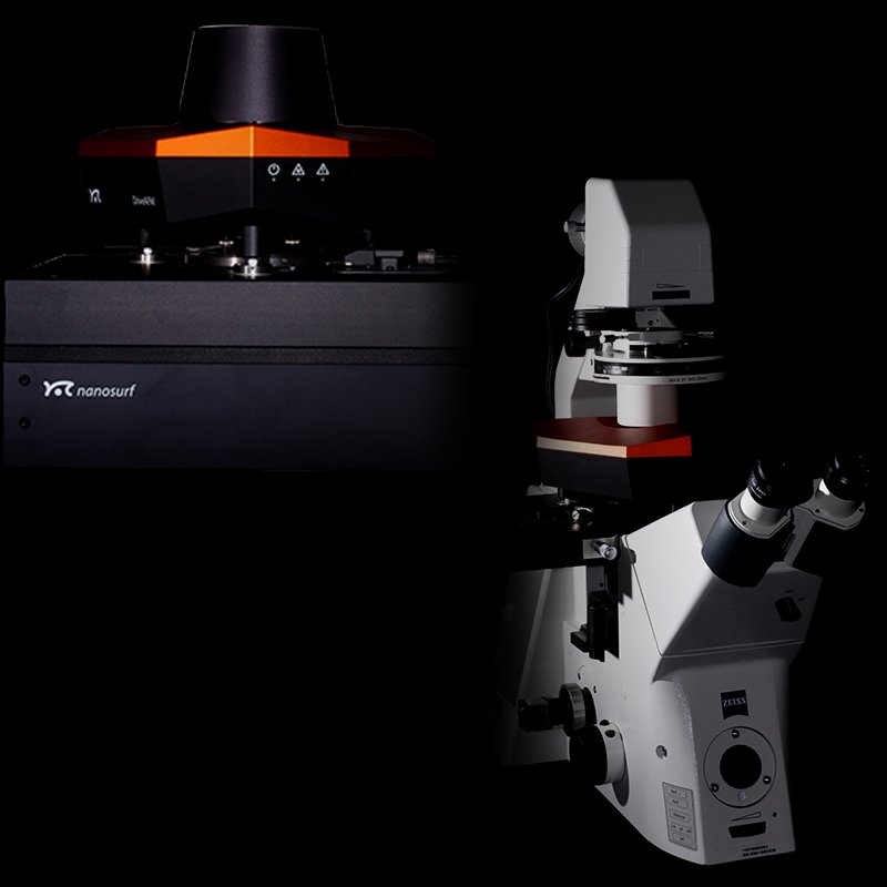
Performance without compromise The DriveAFM is the ultimate tool for nanoscale research. It allows you to explore a variety of phenomena in different domains. With the DriveAFM, you can visualize the DNA double helix, observe the complex patterns of 2D materials and their atomic structure, and measure the electronic properties of semiconductors with high precision. The DriveAFM features CleanDrive, a photothermal cantilever excitation system that ensures optimal stability and performance. It also enables WaveMode, the fastest off-resonance mode on the market. The DriveAFM has a tip-scanning design that accommodates samples of various sizes and shapes without compromising its performance. The DriveAFM, Nanosurf's revolutionary tip-scanning AFM system, empowers researchers with exceptional performance across a diverse range of applications, irrespective of the sample being analyzed. This unique, ultra-low noise architecture provides high-resolution data acquisition, unlocking unparalleled possibilities. It is compatible with high-fidelity small cantilevers that further boost performance. Versatility & Automation No compromising on scan size or setup type. DriveAFM performs in a stand-alone setup as well as integrated with an inverted optical microscope. A full range of imaging modes, the possibility to use small cantilevers, and accessories and options covering virtually all imaginable research needs make DriveAFM future-proof and the best choice for demanding cutting-edge researchers. The DriveAFM comes with full motorization that brings automation features that facilitate setting up and performing experiments. Streamlined Operation with Intuitive Software Nanosurf instruments have always been renowned for ease of use, and DriveAFM brings this to the next level with Nanosurf Studio: a control software designed from the ground up with the purpose of catering to the needs of demanding researchers, and making AFM more intuitively accessible to less experienced users. DriveAFM for Life Science & Biotech DriveAFM plays out all its advantages when it comes to dealing with measurements on biological samples. The full motorization allows you to control the system without interfering with a temperature-controlled environment, and seamless integration of the scan head with an inverted optical microscope allows transmitted light and fluorescence microscopy to be combined with AFM imaging and force spectroscopy. Benefit from correlative techniques and get results that drive forward your research. For Materials Research: The DriveAFM offers a combination of high performance and adaptability for a broad spectrum of applications in materials research. Its design ensures consistent and reliable operation across both air and liquid environments, and effectively handles large and heavy samples. The system is user-friendly, with automation features that streamline the process of conducting measurements on different parts of a sample. Beyond basic imaging, the DriveAFM supports the investigation of a sample's electrical and mechanical properties at the nanoscale. It also accommodates a variety of accessories, enhancing its functionality to include temperature control, magnetic field application, electrical current detection, and the ability to observe dynamic changes in electrochemical processes. For Electronics and Energy Materials: The DriveAFM stands out in the field of electronics and energy storage research, offering a glovebox-compatible solution that ensures precise AFM characterizations in controlled environments, crucial for sensitive samples. Its integrated SMM option and comprehensive NEC features provide a streamlined, user-friendly approach for in-depth nano-electrical characterization of materials, from dielectrics to metals. With minimal sample preparation, quick setup changes, and high-fidelity nanoscale measurements, the DriveAFM facilitates efficient and meaningful insights into the electrical properties of materials, enhancing research outcomes in these advanced sectors. DriveAFM Features: - High-Resolution Imaging: DriveAFM is an optimal tool for high-resolution imaging of biological specimens, capable of capturing details as minute as the structure of single molecules. Its stable CleanDrive photothermal excitation feature ensures reliable performance, even in liquid conditions required to maintain biological sample integrity. Furthermore, CleanDrive paves the path for WaveMode, the most rapid off-resonance imaging mode, enhancing the efficiency of imaging applications. - Most Gentle Imaging with WaveMode: DriveAFM's WaveMode, powered by CleanDrive photothermal excitation, provides unprecedented speed in AFM off-resonance imaging without compromising "gentle touch" crucial for analyzing biological matter. This also enables researchers to investigate samples that before were hardly addressible with conventional AFM imaging modes, such as weakly attached samples or sensitive complexes. CleanDrive ensures the stable and reliable operation. Notably, the chosen 785 nm laser excitation and 840 nm deflection detection wavelengths are biocompatible and non-interfering with biological samples, even facilitating concurrent fluorescence imaging for comprehensive analysis. - Nanomechanics at Your Fingertip: The Drive AFM provides a variety of methods to examine different aspecs of the nanomechanics of your sample, ranging from molecular adhesion or unfolding measurements to the mapping of nanomechanical properties and frequency-resolved dynamic mechanical analysis. The Spectroscopy Plus feature empowers you to delve into force spectroscopy-based nanomechanical experiments. This versatile option opens up a range of possibilities, from basic force-distance curves to more intricate processes involving multiple indentation and relaxation cycles, revealing the viscous nature of your sample. The newly introduced ViscoElastic Analysis option significantly broadens the frequency range for performing dynamic mechanical analysis. This facilitates the investigation of more localized dynamics, such as the relaxation modes of individual filaments within the cytoskeleton. - WaveMode - Off-Resonance Imaging Redefined: WaveMode, a groundbreaking innovation by Nanosurf, revolutionizes off-resonance imaging by transforming the cantilever into a dual-function sensor and actuator. This unique design enables WaveMode to achieve unparalleled imaging speeds, making it the fastest off-resonance imaging mode available. With precise control over the tip-sample interaction, users can adjust the interaction force to meet the specific requirements of the sample, enhancing tip durability and allowing for comprehensive analysis under varying loads. The versatility of WaveMode makes it an essential tool for advanced surface analysis. - Glovebox-Ready: DriveAFM is perfectly suited for operation in a glovebox: full control of all mechanical adjustments via the software, a robust scan head with magnetic cantilever holders for easy operation even when wearing thick gloves. DriveAFM's low-noise, high-fidelity nanoscale platform coupled with the most stringently controlled environment provided by the glovebox hands you everything you need for smooth glovebox operation. Your sensitive samples will be kept in optimal environmental conditions while you perform you AFM characterization. - Advanced Nanomechanical Characterization: The DriveAFM offers a comprehensive set of functionalities to explore the intricate nanomechanical properties of your materials. This includes quantifying adhesion forces, characterizing material response under deformation, and mapping spatial variations in mechanical behavior. The Spectroscopy Plus module empowers in-depth nanomechanical investigations through force spectroscopy. It facilitates a wide range of experiments, from fundamental force-distance measurements to complex protocols involving multi-cycle indentation and relaxation. This enables the characterization of a material's viscoelastic behavior, providing insights into its energy dissipation and deformation mechanisms. The newly introduced Viscoelastic Analysis module significantly expands the accessible frequency range for DMA measurements. This allows for the investigation of highly localized dynamic processes, such as the relaxation behavior of individual components within complex materials. - Nano-Electrical Characterization: DriveAFM's NEC features provide critical insights into the electrical behavior of materials at scales not visible to conventional microscopy techniques. From C-AFM to SMM, the full set of modes is available to analyze your samples. Quick and easy switching of setups for different types of measurements streamlines your measurement work and allows you to focus on what matters: achieving meaningful results. FluidFM: The DriveAFM is ready for research that goes beyond conventional AFM. FluidFM® combines microfluidics with the force sensitivity and positional accuracy of a Nanosurf AFM, unlocking exciting applications in single-cell biology and nanoscience. Conduct original research at the frontiers of science from bacterial adhesion to cell-cell interaction and from spotting to injection and extraction of cells. Benefit from optical, force, and fluidic control, with full software integration on DriveAFM. FluidFM can even be combined with PicoBalance to measure the mass of cells and microparticles picked up by the FluidFM probe.
CATEGORY
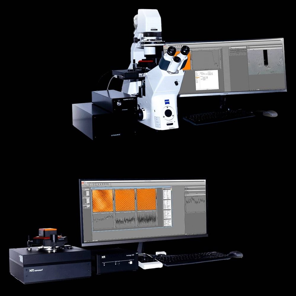
The Flexible Research AFM Biologists, physicists, and material scientists around the globe regard the Nanosurf FlexAFM as the perfect tool for their research needs. This atomic force microscope is designed to be versatile, configurable and user-friendly, making it the preferred choice for hundreds of researchers across various disciplines. Easily integrate it with an inverted optical microscope for biological applications, or set it up in a glovebox for 2D materials research. The FlexAFM offers a wide range of operating modes and accessories designed to enhance and adapt to the particular demands of your research experiments. And as your research evolves, you can upgrade your system without breaking the bank. Versatility and configurability: The FlexAFM is not just a scanning probe microscope. It is a versatile system that can be configured to fit different requirements. Whether you need a stand-alone setup, a glovebox integration or inverted optical microscope compatibility, the FlexAFM can handle it all. With a wide range of options, accessories and operating modes, this AFM is the ideal choice for researchers from various fields of study. The FlexAFM is a trusted, reliable and affordable mid-range AFM system with unmatched flexibility. User-friendliness and ease of use: The FlexAFM combines a compact and lightweight design with robustness and a versatile and user-friendly functionality set. It is easy to switch between different functional sample holders and operating modes. Mounting cantilevers is a quick and simple tasks thanks to the intuitive alignment system. The FlexAFM's camera with integrated top and side view of the samples facilitates sample observation and navigation. The FlexAFM is a flexible and reliable solution that you will love for its easy handling and functionality. Placing a cantilever into the cantilever holder. Alignment grooves precisely position the cantilever. Future-proof investment: The FlexAFM is the smart choice for researchers who already have plans for the future. This versatile and highly adaptable platform will grow in tandem with your research needs. You can easily add new modules and features to the FlexAFM as you explore new applications and techniques. FlexAFM accessories are compatible with the DriveAFM, Nanosurf's most advanced research instrument: when you upgrade your scan head, you will already have your accessories and sample holders ready to go. FlexAFM for Materials Research: FlexAFM shines in materials science applications thanks to its design and powerful suite of modes. Especially when it comes to electrical characterization of sensitive samples, glovebox compatibility and the simple handling make FlexAFM the go-to option for hundreds of materials scientists worldwide. Precision and versatility made cost-effective: The FlexAFM is a tip-scanning atomic force microscope that can handle large samples without compromising on versatility, robustness, or compact system size. Its 24-bit C3000i controller enables high-resolution imaging of even single atomic layers, as well as exploring sample topography spanning several micrometers. The FlexAFM also offers a wide range of accessories and operating modes that enhance its functionality and versatility. With the CX controller upgrade, the FlexAFM can be integrated with external hardware for advanced applications. This is also the first step on the upgrade path to the DriveAFM. Stack of graphene sheets. Image width: 5.11 µm. Glovebox-ready: If you want to protect your samples from environmental contamination but still perform high-quality AFM measurements, the FlexAFM is the ideal choice. The light weight, compact design and magnetic cantilever holder make it easy to operate inside any glovebox. A broad range of nano-electrical modes allow you to explore the properties of new and sensitive materials. The FlexAFM is not only a cost-effective solution, but in many cases it is the best tool for you to achieve your research goals. Nano-Electrical Characterization: The FlexAFM provides a comprehensive collection of nano-electrical modes that provide critical insights into the electrical behavior of materials at scales not visible to conventional microscopy techniques. From C-AFM to SMM, the full set of modes is available to analyze your samples – from 2D materials and semiconductors to metals and piezo-active materials. Quick and easy switching of setups for different types of measurements streamlines your measurement work and allows you to focus on what matters: achieving meaningful results. Conductivity image mapped to the 3D topography of a polished chip with transistor contacts. Image width: 23 µm. Contact potential difference image of graphene surrounded by copper oxide. Image width: 10 µm. FlexAFM for Life Science & Biotech: FlexAFM is the reliable instrument for your bio-AFM applications. The lightweight compact build make it easy and safe to handle with one hand, inverted microscope integration gives you full flexibility to use correlative techniques, and a large selection of sample holders, accessories and add-ons allow you to tailor your FlexAFM research system to fit any avenue of research you are pursuing. A deliberately designed upgrade path to DriveAFM allows you to add benefits at each step of the way. Flexible Design for Life Sciences: Whether you want to study nanomaterials, biomolecules or cells, the FlexAFM can provide you with measurements in liquid environments, where your specimen is in near-physiological condition. You can easily integrate the FlexAFM with most inverted optical microscope from leading manufacturers (Zeiss, Nikon, Olympus, Leica), and benefit from simultaneous optical and AFM data acquisition, in particular when using FluidFM to manipulate cells. Alternatively, you can use the FlexAFM as a stand-alone system for experiments that do not require optical guidance, such as probing the forces interacting within or between single biomolecules or characterizing the structure and properties of biomolecular assemblies or fibrils. FlexAFM Features Motorized Stages FluidFM Python Scripting Nanosurf SMM Option Glovebox-Ready Upgrade Path to DriveAFM FlexAFM imaging modes Standard imaging modes - Static Force Mode - Lateral Force Mode - Dynamic Force Mode (Tapping Mode) - Phase Imaging Mode Electrical properties - Conductive AFM (C-AFM) - Scanning Spreading Resistance Microscopy (SSRM) - Piezoelectric Force Microscopy (PFM) - Electrostatic Force Microscopy (EFM) - Kelvin Probe Force Microscopy (KPFM) - Scanning Microwave Microscopy (SMM) - Magnetic Force Microscopy (MFM) Mechanical properties - Force Spectroscopy (Unfolding and Stretching, Adhesion) - Force Modulation - Force Mapping (Stiffness and Modulus, Adhesion) Lithography and Nanomanipulation Electrochemical AFM (EC-AFM) Scanning Thermal Microscopy (SThM)
CATEGORY
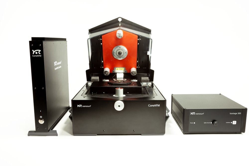
The Best Value Research AFM The CoreAFM was designed to incorporate all the essential elements of an AFM in an easy to use, integrated single unit: a modern flexure-guided scanner, an XYZ sample stage, a camera for sample observation, an active vibration table, and an airflow shielding. The specially designed 24-bit controller ensures precise and reliable control over the scanning process, as well as easy data acquisition and analysis. The CoreAFM system has everything you need to start exploring the nanoworld with AFM; just connect the controller, plug in power and USB, and you are ready to go. Seamlessly extendable functionality The CoreAFM is a versatile and powerful instrument that can handle various applications in both air and liquid environments. Whether you need to perform basic or advanced measurements, the CoreAFM adapts to your needs with its modular design and more than 30 optional modes and functions. Advanced options include sample heating, environmental control, in-plane magnetic field control, and scripting. The CoreAFM is more than just an all-in-one atomic force microscope, it is a complete solution for your nanoscale research and analysis. Easy handling and setup With the CoreAFM, you can explore the nanoscale world with ease and convenience. The CoreAFM is a complete system that requires minimal setup – connect three cables, and you’re ready to go! The user-friendly design allows you to operate the system with one hand. Everything is in its place and within your reach. Basic research AFM with a competitive price tag If you are looking for a reliable and versatile instrument for basic research, the CoreAFM is the right choice for you. This system offers a solid foundation for a multitude of applications in nanoscience and nanotechnology, with the option to expand the feature set as your research evolves. The CoreAFM is a cost-effective solution that delivers high quality without compromising your budget. Whether it is to explore surface morphology or study electrical phenomena, the CoreAFM will help you achieve your objectives with ease and accuracy. CoreAFM for Materials Research CoreAFM incorporates all the essential elements of a research AFM in an easy to use adaptable platform. The precision and performance are well-suited for a wide variety of applications in materials science and related fields. Powerful imaging and precise electrical characterization modes provide you with a robust and versatile research instrument that shines under the pressure of a tight budget. Precision and Versatility The CoreAFM is a powerful and versatile instrument that can handle a wide range of applications in material science and beyond. Whether you are working with polymers, ceramics, metals or 2D materials, the CoreAFM provides you with high-quality data and insights. With more than 30 modes and functionalities to choose from, the CoreAFM allows you to explore different aspects of your samples, such as morphology, mechanical properties, electrical properties and more. Nano-Electrical Characterization With the CoreAFM, you can explore the electrical properties of various materials with unprecedented precision and versatility. Whether you are interested in the conductivity, capacitance, magnetism, or piezoelectricity of your samples, the CoreAFM has a mode for you. From C-AFM to SMM, the CoreAFM covers a wide range of electrical AFM techniques that can reveal the nanoscale secrets of 2D materials, metals, piezo-active materials and semiconductors. The CoreAFM is not just another AFM instrument, it is a powerful tool for electrical nanoscience. Effective and Affordable The CoreAFM combines precision and affordability, making it a smart choice for your research needs. Configure the functionality of your system to perfectly meet your needs, and purchase only what you need. The base configuration includes everything vital for an AFM to perform effectively, and the universe of functionality options offers everything you need to tailor a lean system that supports your research perfectly. CoreAFM for Life Science CoreAFM does not look like the typical bio-AFM, but thanks to its smart engineering can perform very well in certain areas of life-science research. The functionality set includes all features commonly required in life science focused AFM, and the vital aspect of microscope integration is available through the DIMO. CoreAFM is the only AFM in its class with versatility covering even biological applications. It’s also the most affordable path to FluidFM functionality. CoreAFM Features Compact All-in-One Device Nanosurf SMM Option Python Scripting Variable Magnetic Field Generator (in-plane) FluidFM Digital Inverted Microscope Option CoreAFM imaging modes Standard imaging modes - Static Force Mode - Lateral Force Mode - Dynamic Force Mode (Tapping Mode) - Phase Imaging Mode Electrical properties - Conductive AFM (C-AFM) - Scanning Spreading Resistance Microscopy (SSRM) - Piezoelectric Force Microscopy (PFM) - Electrostatic Force Microscopy (EFM) - Kelvin Probe Force Microscopy (KPFM) - Scanning Microwave Microscopy (SMM) - Magnetic Force Microscopy (MFM) Mechanical properties - Force Modulation - Force Spectroscopy (Stiffness & Modulus, Adhesion, Unfolding & Stretching) - Force Mapping (Stiffness and Modulus, Adhesion) Lithography and Nanomanipulation Electrochemical AFM (EC-AFM) Scanning Thermal Microscopy (SThM)
CATEGORY
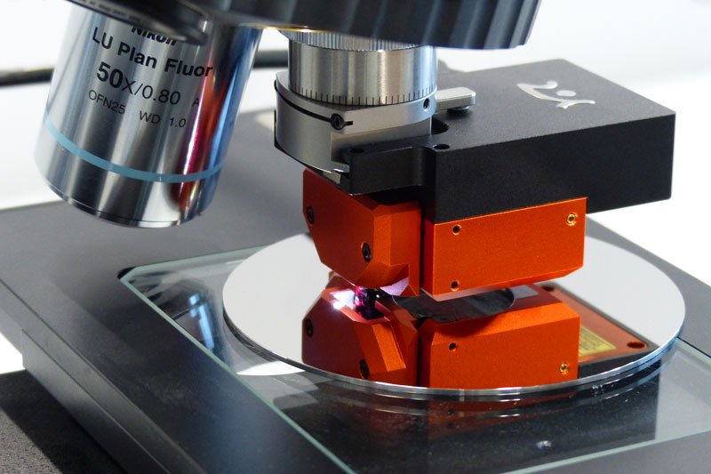
Extend the Resolution of Optical Microscopes The Nanosurf LensAFM is an atomic force microscope that picks up where optical microscopes and profilometers reach their resolution limits. It is mounted like a normal objective lens, thus extending the resolution and measuring capabilities of these instruments. The LensAFM not only provides 3D surface topography information, but can also be used to analyze various physical properties of a measurement sample. Seamless Integration In an ever-increasing number of situations, researchers are looking to combine optical and atomic force microscopy techniques. The ease of use, screening capability, and minimal sample preparation requirements of optical microscopes are almost unparalleled. However, when the resolution of a 100x objective is not sufficient to examine small features beyond the instrument's resolution, the LensAFM comes into play. Its exceptionally small design and clever mounting mechanism mean you only need to rotate the turret on your optical microscope or profilometer and run the scan. Enhance Your Optical Microscopy with AFM for Advanced Insights Since the resolution of optical microscopy is limited by the wavelength of light, there is a barrier in the resolution you can achieve with your optical system. In an ever-increasing number of applications, this calls for the combination of optical and atomic force microscopy. In addition, AFM overcomes problems characterizing transparent samples or samples otherwise difficult to assess optically. But not only the coarse topography of a sample is of interest: AFM also allows knowledge of other material properties to be acquired, e.g. surface roughness, hardness variations, magnetism, or electrical conductance/resistance. Precision Mounting Featuring a quick-release mechanism, the LensAFM is easily mounted and unmounted from the turret. Its kinematic mounting guarantees replacement with better than 10 µm accuracy. Furthermore, alignment grooves on the chip mount ensure that the tip of the next cantilever is within 4 µm of the same position, allowing you to find the same feature again, even after a cantilever exchange. Time-Saving Features The LensAFM not only enhances the precision of measurements but also saves time. Thanks to the alignment grooves, there's no need to perform a laser alignment on the cantilever, which streamlines the process and saves additional time. This makes the LensAFM an invaluable tool in any research setting that requires the detailed analysis provided by atomic force microscopy. Workflow Efficiency The LensAFM integrates perfectly into your existing workflow. Once mounted on your optical microscope's turret – just like a regular objective lens – you can screen the sample with optical methods to find areas of interest. Subsequently, these areas of interest are easily found again using the integrated 8x optical lens, and then you can perform your AFM measurement for higher resolution 3D information. This approach allows you to work in the way you are used to but with a significant boost in resolution and capabilities. LensAFM Features Universal Compatibility: Mountable on virtually any optical microscope or 3D optical profilometer Easy Positioning: Sample positioning using the optical microscope’s position manipulators Optical View: Equipped with an 8x objective lens for a clear view of sample and cantilever Large Z-Range Allow: The large Z-range allows measurement of high structures Automatic Cantilever Approach: Bring your sample into optical focus and the LensAFM does the rest Easy to Use: From imaging to electrical properties and spectrocopy LensAFM Imaging Modes Standard imaging modes - Static Force Mode - Lateral Force Mode - Dynamic Force Mode (Tapping Mode) - Phase Imaging Mode Electrical properties - Conductive AFM (C-AFM) - Electrostatic Force Microscopy (EFM) - Magnetic Force Microscopy (MFM) Mechanical properties - Force Spectroscopy - Force Modulation - Force Mapping Lithography and Nanomanipulation
CATEGORY
