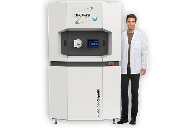
ChipMill A fully integrated solution for millimeter-scale delayering of both logic and memory semiconductor devices. The ChipMill integrates signals from multiple detectors via an artificial intelligence feedback control algorithm to adjust milling parameters in real time. The result is the precise removal of device layers and a highly planar surface. The Model 1064 ChipMill is a fully integrated solution for large-scale delayering – up to a 10 x 10 mm milling area – of both memory and logic semiconductor devices. Compared to all other methods, the ChipMill produces the flattest surface over the largest area. The ChipMill’s artificial intelligence automatically adjusts milling parameters to yield nanometer flatness within the prepared area. The ChipMill reduces time and costs associated with research and development, manufacturing, quality assurance, and failure analysis by yielding unsurpassed results. As the demand for semiconductor devices grows, device sizes decrease, and architecture becomes more complex, the ability to perform controlled delayering during all phases of the product life cycle is essential. - Nanometer flatness of the prepared area - Milling area up to 10 x 10 mm - Automated sample height detection - User-friendly interface for the setup of milling parameters and display of images and analytical data - On-device touchscreen for managing sample insertion and removal - End-pointing by time, chip structure, or chemical composition - Components: - Ion source - Optical camera - Electron beam column - Secondary electron detector (SED) - Backscattered electron (BSE) detector - Energy dispersive X-ray spectrometer (EDS)
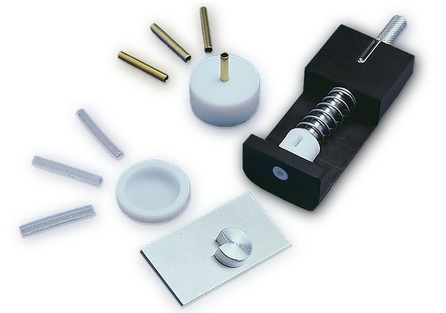
Includes all the components required to produce high quality cross-section specimens for transmission electron microscopy. Widely used cross-section specimens include semiconductor devices (which often have multiple layers and, thus, multiple interfaces), specimens with thin film layers, and composite materials. For the study of interfaces by transmission electron microscopy, it is critical to align the interface of interest parallel to the incident electron beam. One method for preparing such samples is to fabricate cross-section specimens. - Stacks and holds cross-section specimens - Aligns areas and interface of interest - Produces consistent glue layer thickness - Resulting stacks are readily cut by the Model 170 Ultrasonic Disk Cutter - Fabricate self-supported 3 mm diameter disk specimens
CATEGORY
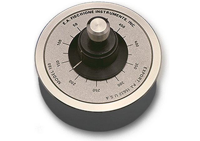
Specimen Grinder. Mechanically prethins specimens for transmission electron microscopy. Produces uniform thickness specimens with parallel sides. Greatly reduces the time spent during the final preparation process of ion beam milling. For transmission electron microscopy, the quality of the initial disk determines the quality of the final specimen. The disk can be mechanically ground using the Model 160 Specimen Grinder to a precisely controlled thickness. Use the Model 200 Dimpling Grinder for final thinning. - Accurate and dependable - Precisely controlled - Specimens up to 18 mm diameter - No additional force needed - Large diameter provides excellent stability - Platen transferable to the Model 200 Dimpling Grinder
CATEGORY
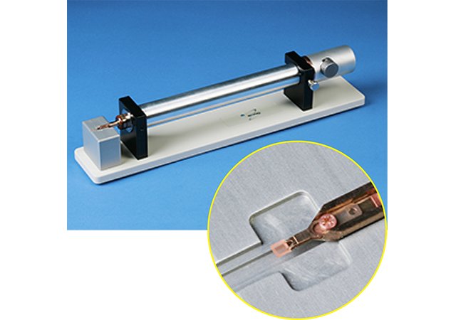
Ultra-Narrow Gap Tomography Holder. A holder capable of tilting up to 90° while providing a maximized field of view for transmission electron microscopy. The Model 2030 Ultra-Narrow Gap Tomography Holder is capable of tilting up to 90° while providing a maximized field of view. The holder is suitable for ultra-narrow gap (< 3 mm) pole-piece geometries. The Ultra-Narrow Gap Tomography Holder accepts a 1.5 mm square or round TEM specimen or grid into a cartridge, securing it with a single clamp. Items included are a specimen loading stand, a Model 9010 Vacuum Storage Container, a loading fixture, a clamp tightening tool, copper grids, tweezers, and a spare parts kit. The holder: - Specimen loading station allows easy positioning and clamping of the specimen into the cartridge - Has a streamlined specimen clamping mechanism eliminates the shadowing associated with most holders at high-tilt angles - Has a retractable clamp that evenly distributes force on the specimen - Accommodates specimen thicknesses up to 100 μm - Is compatible with the TEM’s touch alarm, which stops goniometer movement in the event that a pole touch occurs The holder is available for both JEOL and Thermo Fisher Scientific microscopes.
CATEGORY
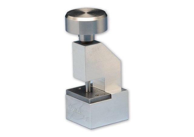
Produces high quality disk specimens from thin metal foils. A precision ground punch and die plate eliminate specimen stress and distortion. An ideal accessory to the Model 110 Automatic Twin-Jet Electropolisher. The specimen punch is available with a standard base or with a wide base, which provides extra stability when producing thin foils. A precision ground punch and die plate eliminate specimen stress and distortion of the disk specimen. A spring-loaded return plunger keeps the disk specimen on the die plate surface for convenient handling. Available in standard sizes of 1 mm, 2.3 mm, and 3.0 mm; other sizes available upon request
CATEGORY
