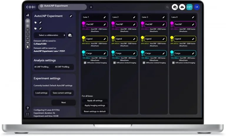
Experiment setup One-click channel mapping Calibration & Sample Check Field of view selection Automatic 4-lane acquisition Summary, LNP montage & Data export Experiment setup The first step of AutoLNP is to input your experiment settings, such as the experiment name and collaboration and any relevant tags. Optimized acquisition and analysis settings for the LNP Profiler kit are set as default for users, including channel names, laser power, number of frames, exposure time, and optimal analysis workflow for automated and accurate end to end workflow. Settings can be adjusted to fit different experimental settings, easily saved and applied across all lanes. One-click channel mapping Channel mapping is an essential step towards generating accurate multicolor super-resolution data using a calibration slide. AutoLNP makes channel mapping even easier with a one-click procedure that only takes around 2 minutes using a bead slide. The channel mapping tool optimizes the 560 nm and 640 nm laser powers, acquiring as many fields of view as necessary to complete the calibration. Calibration & Sample check System calibration is a critical step that uses both Autofocus and AutoTIRF tools to automatically find the imaging surface of the LNP Profiler Assay Chip, lock the focus on the chip, and determine the optimal TIRF illumination angle for higher imaging quality. Sample pre-check is an optional tool that quickly scans each lane of the chip, providing representative images of each lane. This helps confirm sample calibration, optimal focus and illumination, as well as sample density and particle staining. Field of view selection Field-Of-View (FOV) selection can be performed either by choosing the number of FOVs to be imaged at the top of each lane, or manually selecting FOVs within each lane's imaging area. The tool includes estimated time indicators to predict experiment duration and a color-coded system to easily visualize the position of the currently selected FOV, those already acquired, or those used for calibration to avoid repeated imaging. On a Nanoimager microscope, AutoLNP images a FOV of 50 µm by 80 µm, with each imaging area surrounded by a bleaching safe zone so that neighboring FOV are not affected by stray laser light. Automatic 4-lane acquisition A live camera view shows blinking fluorophores during super-resolution imaging. The software provides simultaneous real-time image analysis and information including live localizations from the blinking fluorophores and overall experiment progress. After being acquired, each dataset is automatically uploaded to CODI, thus, AutoLNP also provides information on the FOVs being imaged, those being uploaded to CODI, those being analyzed, and the FOVs where analysis is completed and the number of particles/clusters found. Summary, LNP Montage & Data export The acquisition summary page provides a visual overview of datasets, including imaging settings, FOVs for imaging or calibration, links to CODI analyses, and cluster counts. It shows a mosaic of randomly selected LNPs by positivity class. Users can export data via three tools: the LNP montage tool (snapshots by signal and size), a two-page summary report (population stats, size, PDI, biomarker positivity, cargo/ligand abundance), and a CSV file for download and sharing. Does AutoLNP have manual controls as well? AutoLNP introduces great new algorithms like AutoFocus, AutoTIRF, and automated acquisition across lanes. You can manually set the illumination (TIRF) angle if desired, and you can adjust the focus on a per-lane basis prior to starting the automated acquisition, so you can be sure to have the imaging results you want, all while being able to step away from the microscope for imaging.
This kit contains two single-domain antibodies (sdABs) for DNA-PAINT as well as the necessary imager strands and buffers that are required for sample preparation and imaging. Each sdAB can be coupled to a single DNA-PAINT docking site and two sdABs can bind one primary antibody, resulting in two docking sites per primary antibody. The reagents are tested and optimized for the Nanoimager. Simply add your primary antibodies of interest and you are ready for 2 color DNA-PAINT imaging.
CATEGORY
This kit (MASSIVE-sdAB ONI 2-PLEX Kit) includes two single-domain antibodies (sdABs) for DNA-PAINT, along with imager strands and buffers for sample prep and imaging. Each sdAB binds a DNA-PAINT docking site, with two sdABs per primary antibody. Optimized for ONI microscopes—just add your primary antibodies for 2-color DNA-PAINT imaging.
CATEGORY
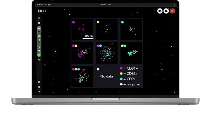
When combined with our newest software offering, AutoEV, your system will be calibrated and optimized to acquire and analyze a 4-lane chip automatically and deliver a comprehensive report of EV size and positivity for each individual lane in 90 mins.
CATEGORY
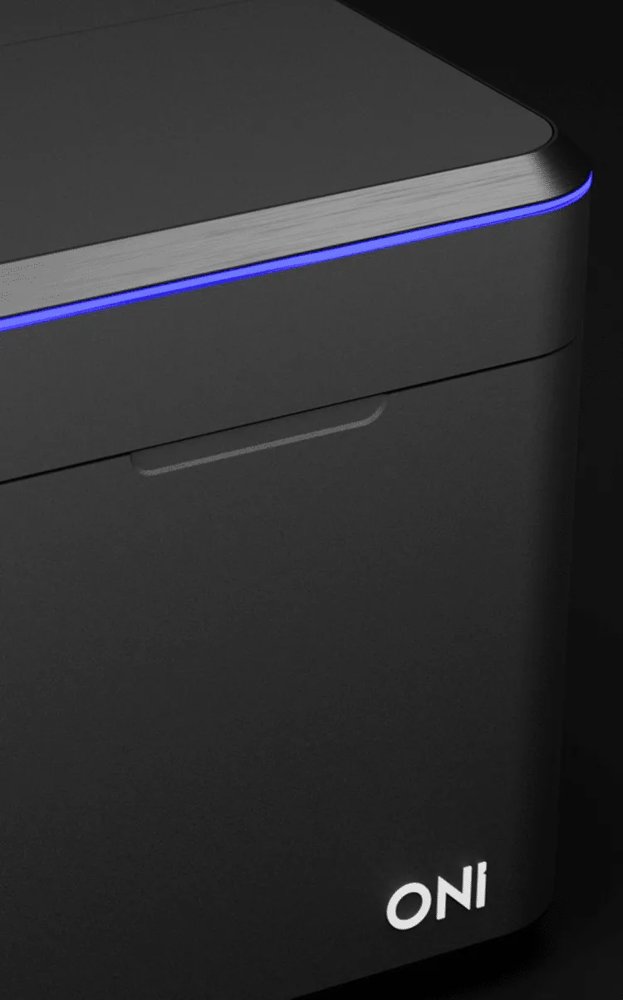
Simple. Engineered. Dynamic. Powerful. A new era in super-resolution imaging Meet Aplo Scope – ONI’s new single-molecule super-resolution microscope designed for effortless molecular imaging. Perfect for researchers of all levels, Aplo Scope combines advanced imaging, precise laser control, and intuitive software to deliver high-quality results, from research on the foundations of life to drug discovery, biomarker research, and disease studies. With its compact design, Aplo Scope transitions seamlessly from live-cell to super-resolution imaging down to 15 nm resolution, without complex setup.Its unparalleled ease of use and integration with the Aplo Platform make it ideal for uncovering molecular interactions and driving breakthroughs. Simple Designed to simplify complex microscopy with one easy-to-use tool. Start imaging quickly, generate data with ease, and unlock answers quicker than ever before. Engineered Robustly engineered for stability, consistency, and reliability, to maximize reproducibility across experiments and users. Dynamic Seamlessly transition between live-cell imaging and super-resolution without reconfiguration for unmatched flexibility in one platform. Powerful Compact footprint, yet powerful enough to enable complex scientific inquiry. Aplo Scope is designed to fit into your lab, not the other way around. The new and groundbreaking SMLM microscope More Channels, More Insights Flexibility Redefined Maximize Throughput Homogeneous Illumination More Channels, More Insights Multicolor imaging reveals biological context Achieve maximal spectral separation with minimized cross-talk and background thanks to an optimized filter set including single, dual, and quad options. Using only 4 lasers, you can generate detailed multi-color panels of 5 or more labels to elucidate complex protein and particle interactions that span both large pan-cellular structures and nanoscale molecular features. Leverage the rapid laser switching with multiband filters for nearly simultaneous multi-channel imaging. Flexibility Redefined Live cell to super-resolution imaging Effortlessly capture dynamic cellular events in real-time, then zoom into molecular details with super-resolution precision — all within a single, intuitive platform designed to enable your cutting-edge science. From low-power live-cell imaging to high-power super-resolution imaging, the Aplo Scope eliminates the need for hardware reconfiguration. Aplo Scope includes unique illumination stability, even at extremely low power (<0.01 kW/cm2), to minimize phototoxicity in extended live cell imaging sessions. At the other end, high laser powers enable rapid, super-resolution imaging to uncover molecular level details. Maximize Throughput Expansive field-of-view Aplo Scope contains a 110 by 110 micrometres field-of-view, in both diffraction-limited and single-molecule localization microscopy. The expansive field-of-view significantly increases the chances of capturing rare events and elusive phenotypes. Detect ultra-rare cell populations and particle biomarkers with unmatched throughput, accelerating groundbreaking discoveries and driving transformative breakthroughs. Homogeneous Illumination Variation that reflects biology Aplo Scope produces a consistently flat, symmetric illumination across the entire field of view, vital for statistical and comparative intensity analysis. With consistent resolution and precision edge to edge, every frame produces more data, maximizing throughput of critical and sensitive assays. This enables quantitative molecular studies to better understand crucial mechanisms in disease and drug discovery studies. Key features of Aplo Scope Next-gen lasers Power density continuously adjustable from 0.01 through 3.325 kW/cm2 Light engine Packed with technology to enable diffraction-limited and SMLM imaging Illumination Illumination angle control from Epi to HILO and TIRF. Consistent symmetric illumination across the field-of-view Temperature control Precise temperature control up to 40°C Filter Wheel Four laser channels can be interlaced with any combination of emission filters to perform multicolor imaging, easily generating 5-color overlays Camera Sensitive next-gen CMOS camera for low noise, high-speed imaging Focus Z-lock Rapid and precise focus with locking technology to minimize Z-drift Field-Of-View 110 by 110 micrometres in both diffraction-limited and single-molecule microscopy Stage Fully automated piezo stage fits most microscopy slides and plates Mechanical stability Integral dual-stage isolation system minimizes vibration Sample enclosure Integral lid interlock ensures user safety, class 1 laser instrument Aplo Scope Aplo Scope is the flagship innovation behind the Aplo Platform: solid performance in a versatile, easy-to-use microscope. Experience a range of imaging modalities with this highly compact microscope designed to simplify multicolor super-resolution microscopy. Aplo Scope allows you to start imaging right away, no dark room or optical table is required. Its integrated software enables fixed or live imaging anywhere, and seamless up to 5 color multimode images! How much training is needed to get started using Aplo Scope? With unparalleled ease-of-use and simplicity, Aplo Scope has been designed to eliminate barriers to entry into the traditionally complex world of super-resolution microscopy and single-molecule imaging, and allow users of any skill level to utilize the instrument within one day of training. What are the dimensions of Aplo Scope? The footprint of the microscope body is as small as a standard laptop 220 mm (w) x 421 mm (d) x 242 mm (h), the light engine measures 215 mm (w) x 420 mm (d) x 456 mm (h) and looks like a computer tower. They’re attached to each other by a 1.5 m long cable and, importantly, can be used on any bench or desk without needing a dark room or optical table. Is Aplo Scope compatible with third-party kits and data analysis tools? Yes, samples can be stained with any super-resolution microscopy-compatible dye and imaged using Aplo Scope. However, this would require manual acquisition and analysis within CODI, or a third party application. ONI offers end-to-end kits where sample preparation, image acquisition, analysis and reporting are all integrated into a workflow, making imaging a seamless and more automated experience.
CATEGORY
