SPRm 200 System is the world’s first commercial Surface Plasmon Resonance Microscopy (SPRM). It combines the high spatial resolution of optical microscopy with the powerful sensing capability of SPR, making it possible to measure the binding of molecules label free with membrane proteins on single intact cells for the first time. Cells are incubated on a traditional SPR sensor surface, the analyte molecules are introduced with a flow system to allow the binding of the molecules with membrane receptors on the cells, and the binding kinetics is obtained for each of the cells on the surface from the single-cell sensorgrams. The association constant ( ka), dissociation constant ( kd), as well as the equilibrium constant (KD) are obtained. Unlike traditional SPR systems, SPRm 200 provides high spatial resolution and simultaneous optical microscopy capability, which are necessary to resolve and measure molecular binding taking place on the surface of individual cells. With SPRm 200, the SPR response is mapped in a 600 μm x 450 μm image and the binding affinity and kinetics information is collected at each pixel of the image. By providing a sensorgram at each pixel of the image, the instrument offers spatial mapping of the biding activities over any given sensing area, yielding much more information than the conventional SPR systems. A particularly unique application of SPRM for studies of biomolecular interactions with multiple cells is exemplified by the studies of binding between glycoproteins at cell membranes and their ligands. Glycoproteins, which are important for cell recognition and communications, are membrane proteins with sugar groups extended into the extracellular space of the cells 1. The interaction of wheat germ agglutinin (WGA, 36k MW), a lectin that binds sugar groups and can recognize N-acetylglucosamine (GlcNAc), on live and fixed Barrett’s esophagus-derived CP-D (CP-18821) cells was investigated in this note. WGA exposure to these cells resulted in an increase of SPR response in the SPR image, which indicates the presence of sugar residues on the glycan chain of the membrane protein 2.Fig.1 shows the application of SPRm 200 to measure the binding of lectin with glycoproteins on the membranes of individual live cells. The bright-field optical microscopy of SPRm 200 reveals the individual cells (Fig. 1A), while the SPRM image (Fig. 1B) provides valuable binding signal on each of the cells at a specific concentration, from which SPR sensorgrams for the individual cells are obtained (Fig. 1C) for wheat germ agglutinin (WGA). For this study, the cells were incubated on the sensor surface at 37 °C with 5% CO2 and 70% RH. The cell surface was regenerated with 50 mM N-acetylglucosamine (GlcNAc). Only two concentrations (87nM & 175nM) were tested in this set up due to cell apoptosis. Fig 1. Bright field image of live CP-D (A), SPR image of CP-D binding to glycoprotein receptors (B), SPR sensorgrams of selected cells (C). From the eleven regions (live cells) selected for analysis, the average ka, kd and KD were 1.14 x10 4 M-1 s-1, 3.42 x 10 -4 s-1 and 38.7 (+/-24) nM, respectively. Information, descriptions, and specifications in www.biosensingUSA.com
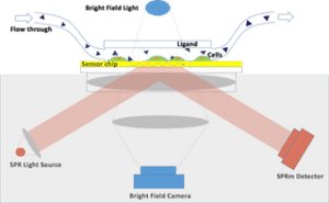
The SPRm 200 from Biosensing Instrument Inc. is a system that seamlessly combines both optical bright-field and SPR microscopies thus enabling the unique in vitro binding kinetic measurement of individual cells in their native environment. The architecture of the system consists of using the SPR phenomenon to measure binding kinetics while also simultaneously permitting use of bright-field microscopy to study topographical and phenotypical changes in the sample (Fig. 1). Typically, the cells are cultured and fixed on a poly-L-lysine coated SPR sensor chip. The integrated optical microscope allows for visualization of the sample with x10 magnification and 1 um resolution. The SPRm 200 exceeds the performance of homemade systems by demonstrating a much larger field of view, simultaneous bright-field and SPR microscopies, and greater stability. SPRm 200 has the resolution to observe sub-cellular binding activities (Fig. 3). In conclusion, SRPm 200 is a novel instrument that seamlessly integrates bright-field and SPR microscopies into one system. This instrument enables the kinetic binding study of multiple individual cells as well as the localized responses on a single cell.
CATEGORY
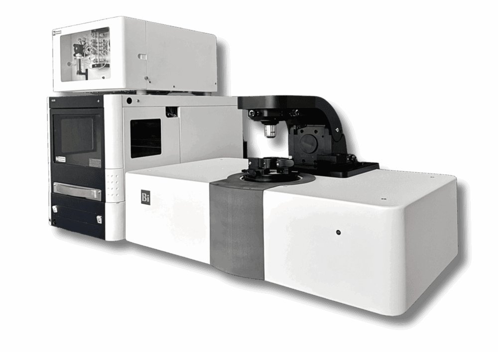
The Surface Plasmon Resonance (SPR) Microscopy system, which combines optical microscopy with SPR, is a powerful and label-free technology for measuring the binding affinity and kinetics of membrane proteins directly on whole cells in vitro. This integrated approach enables the simultaneous analysis of phenotypic changes via brightfield imaging and molecular interactions through SPR, offering a comprehensive view of cell-surface binding events. SPR Microscopy has been replacing radioligand binding assays and has become a go-to platform in both pharmaceutical and academic research for studying the interactions of small molecules, peptides, antibodies, and proteins with membrane targets such as GPCRs, ion channels, and other difficult-to-purify membrane proteins. It supports measurements on both fixed and live cells, whether adherent or in suspension. Thanks to its high spatial resolution (~1 μm), the technology also enables real-time monitoring of nanoparticles, virus and bacterial binding activity, expanding its applications in infectious disease and microbiology research.
CATEGORY
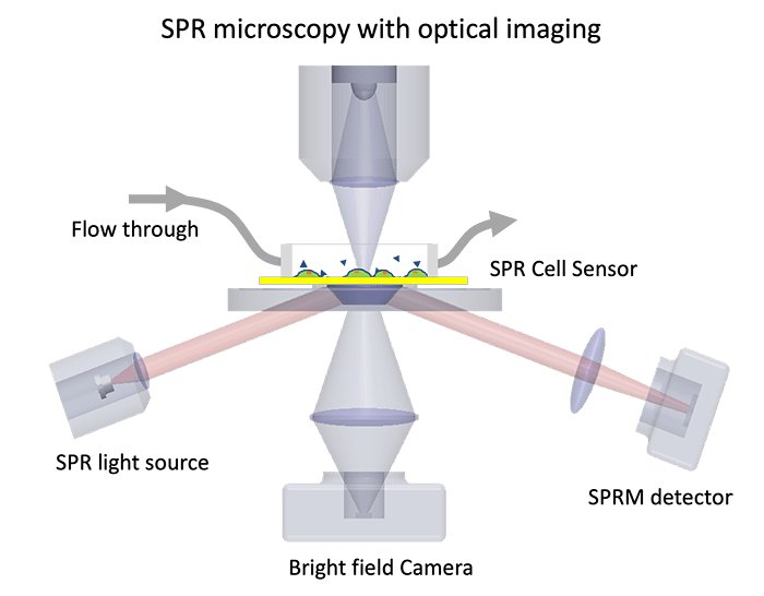
SPR microscopy integrates optical imaging and SPR technology into one system. It provides high-quality data on binding kinetics and affinity of cell membrane proteins and spatial visualization of cellular structures. Real-time interactions of the drug and membrane protein can be measured in its native state without needing to extract proteins from the cell. Because of its unique measurement of cell membrane proteins on the cell, SPR microscopy has become a powerful tool that provides binding information about various drug compounds and drug efficacy. Antibody-interaction studies are also used to investigate disease pathways and targets for potential drug therapies, and much more. In addition, the system measures nanometer scale binding activities needed for the study of bacteria and virus interactions and the development of new methods for nanoparticle drug delivery.
CATEGORY
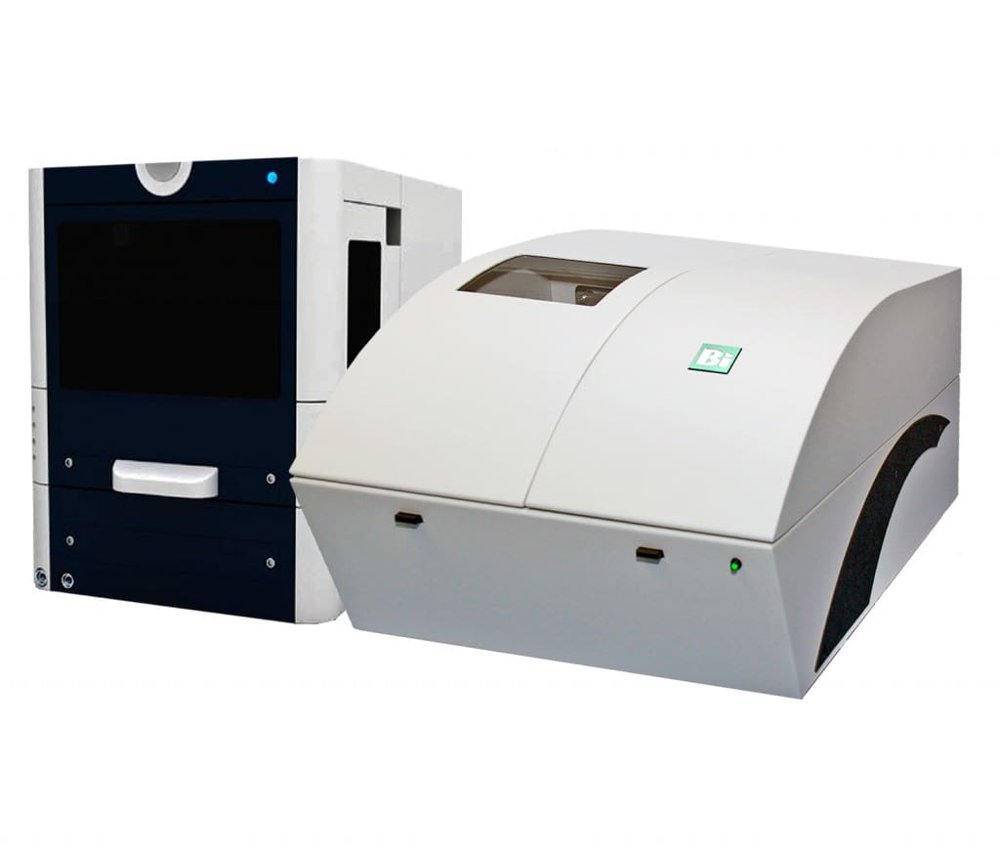
The 5-channel SPR instrument, when paired with the BI Autosampler, offers an ideal solution for high-throughput assay development, combining efficiency with low operating and maintenance costs. Featuring advanced BI-DirectFlow™ technology—integrated into the BI-4500 system—it ensures precise sample delivery for exceptional binding kinetic measurements. This innovative flow system, together with temperature-controlled SPR detection, provides highly sensitive and reproducible binding responses, even for low immobilization levels and small molecule targets (<100 Daltons). The system also supports compatibility with electrochemistry and chemical vapor analysis modules, enabling integrated EC-SPR and gas-SPR measurements. These capabilities make it especially well-suited for a wide range of biosensing applications.
CATEGORY
