By Gatan
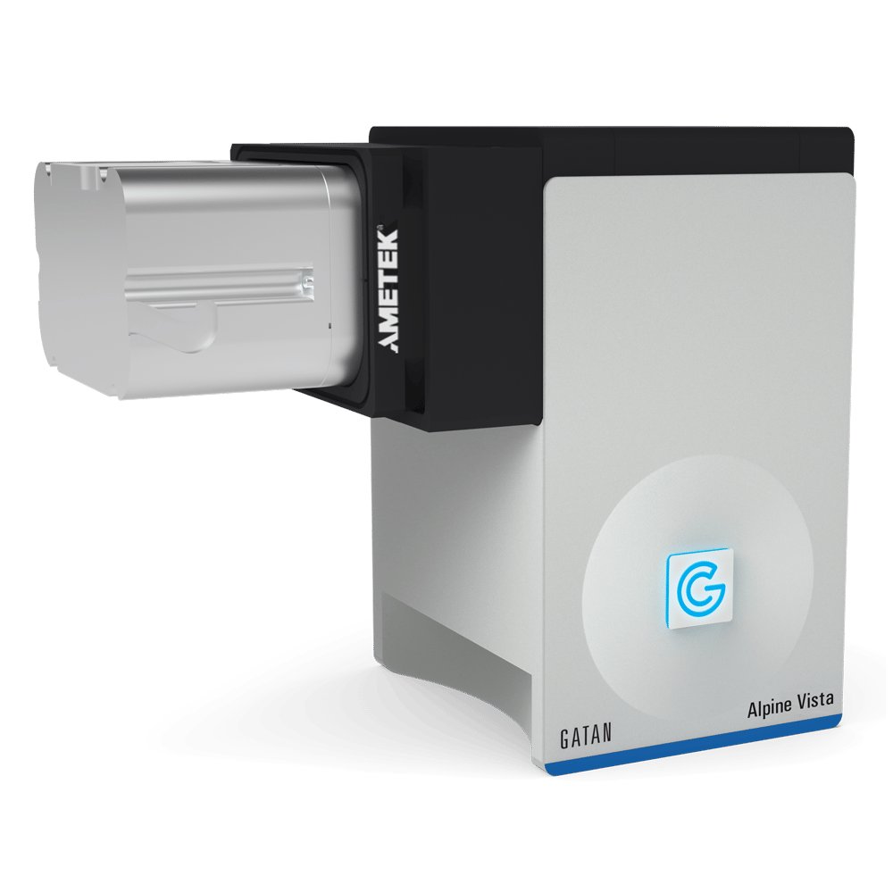
Models 1124, 1125. The world’s first direct detection cameras to democratize cryo-EM. Alpine®is the world’s first direct detection camera series to democratize cryo-electron microscopy (cryo-EM) imaging. By enabling the acquisition of high-resolution structures on 80 – 200 keV microscopes, the Alpine series allows routine cryo-EM research on more affordable, workhorse instruments and expands its usefulness to the larger research community. Two models available to match the field of view with your application needs: Alpine Vista (1125) – Maximizes imaging throughput for your high-performance microscopes with a large FOV of 5,760 x 4,092 pixels. Delivers a detective quantum efficiency (DQE) up to 2.4-times the performance of scintillator-based cameras. Makes the 2D classification of small molecules possible while improving macromolecule reconstructions. Streamlines your workflow and detects failures early to produce better structures, faster. Real-time counting ensures there is no compromise between data collection speed and quality.
Models 1124, 1125. The world’s first direct detection cameras to democratize cryo-EM. Alpine®is the world’s first direct detection camera series to democratize cryo-electron microscopy (cryo-EM) imaging. By enabling the acquisition of high-resolution structures on 80 – 200 keV microscopes, the Alpine series allows routine cryo-EM research on more affordable, workhorse instruments and expands its usefulness to the larger research community. Two models available to match the field of view with your application needs: Alpine (1124) – Turns 80 – 200 keV screening microscopes into data-collection microscopes with single-electron counting capabilities. Delivers a detective quantum efficiency (DQE) up to 2.4-times the performance of scintillator-based cameras. Makes the 2D classification of small molecules possible while improving macromolecule reconstructions. Streamlines your workflow and detects failures early to produce better structures, faster. Real-time counting ensures there is no compromise between data collection speed and quality.
CATEGORY
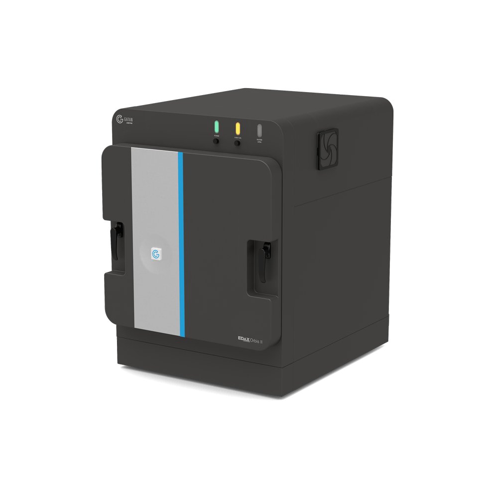
Model 1140 Delivers non-destructive, high-performance elemental analysis with unmatched flexibility and ease of use. The EDAX® Orbis™ II micro-x-ray fluorescence (micro-XRF) system delivers non-destructive, high-performance elemental analysis with unmatched flexibility and ease of use. Engineered for excellence across the full spectrum of micro-XRF applications, the Orbis II empowers users to analyze everything from the smallest particles to large, complex samples—quickly and accurately. Experience the next level of micro-XRF analysis with the EDAX Orbis II—where precision meets performance. - Custom x-ray window technology enables the detection of light elements - Proprietary coaxial light and x-ray optics precisely targets specific regions – eliminating x-ray interference from adjacent areas - Maximizes throughput by collecting spectra 2.5x faster and spectral maps 3x faster - Streamlines workflows with programmable autorun routines to boost productivity and ensure consistent results
CATEGORY
Micro X-ray Fluorescence EDAX Orbis II Micro-XRF System Delivers non-destructive, high-performance elemental analysis with unmatched flexibility and ease of use.
CATEGORY
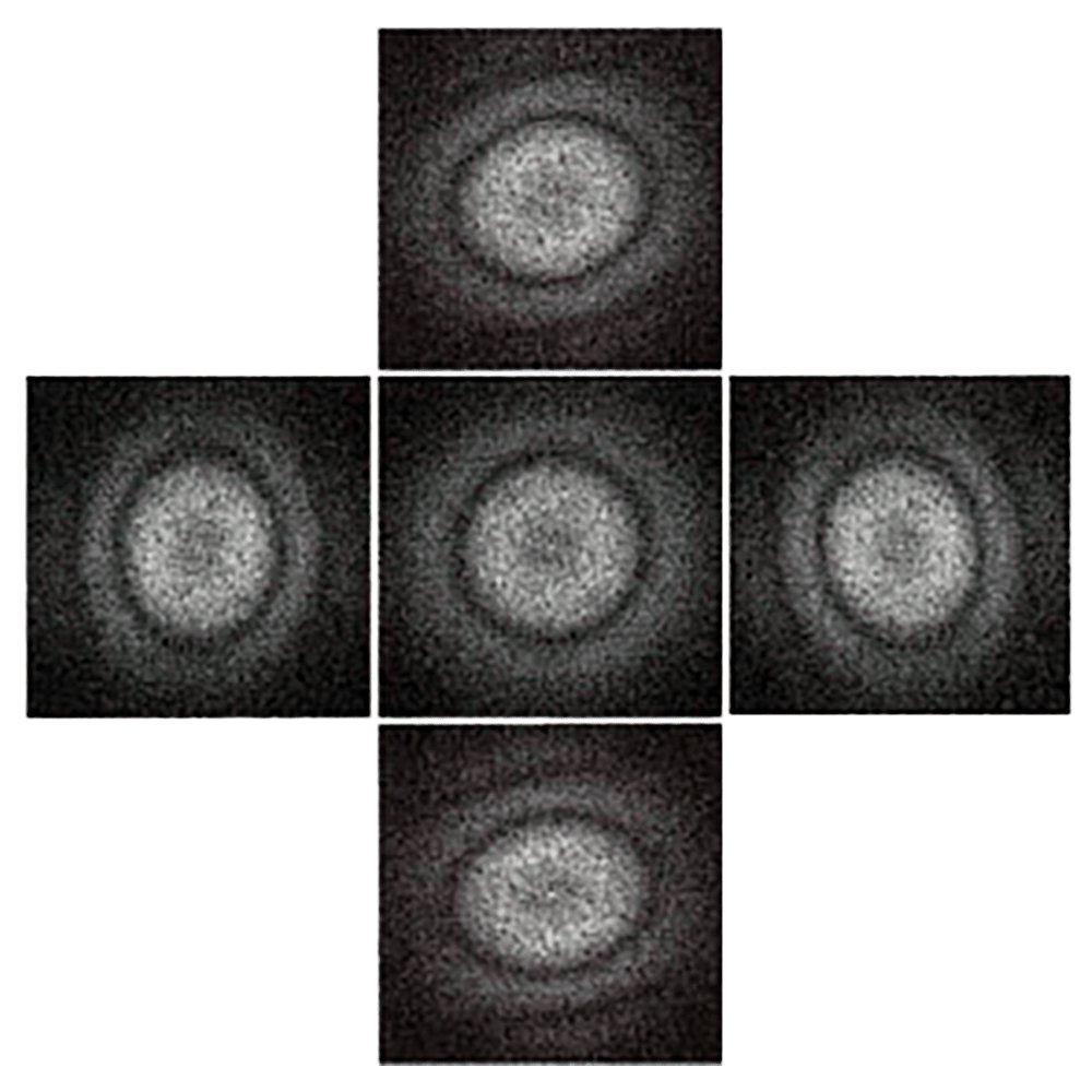
Stage and optics control so you can seamlessly stitch images together. Advantages By controlling the microscope stage or imaging optics, DigitalMontage®software enables an array of images to be acquired and stitched seamlessly together. The resulting montage shows specimen structure at a resolution that would be impossible to collect even with cameras possessing ten times more pixels. Acquisition To offset the field of view of the camera, DigitalMontage software can use either the microscope stage or the image-shift deflectors. At high magnifications it may be easier to stitch the images together if the image-shift deflectors are used to offset the field of view. At low magnifications the stage enables a larger area of the specimen to be covered and minimizes the effects of any optical distortion present in the system. Stitching When stitching the images together DigitalMontage software automatically adjusts the intensities of the images so as to minimize or eliminate the presence of seams in the result. If you should encounter a case which is difficult to automatically stitch, you can choose to refine the positions of the images after the automatic determination, or to completely manually position them. The software will then stitch the images together and adjust the intensities as in the fully automatic case. Building upon experience gained from the past, this version of DigitalMontage has been completely re-written and integrates seamlessly with the rest of the DigitalMicrograph® software.
CATEGORY
