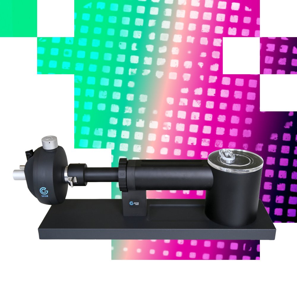
Cryo-EM solution for analytical 4D-STEM characterization of beam-sensitive samples Cryo-Transfer Holder for Tescan TENSOR™ The Tescan TENSOR™ cryo-EM solution extends the unique and accessible analytical capabilities of TENSOR™ to cryo-STEM, cryo-EDS, cryo-3D ED, and cryo-4D STEM analysis of beam-sensitive materials by combining a cryo-transfer holder with low-dose procedures in Tescan Explore™ software. Reduced Beam Damage for Advanced Materials Characterization The single-tilt Gatan Elsa™ cryo-transfer holder, fully adapted for compatibility with Tescan TENSOR™, is designed for stable, high-resolution cryo imaging at liquid nitrogen temperature, with a hold time of more than 8 hours. The cryo-transfer holder is part of the TENSOR™ cryo-EM solution, which combines cryogenic sample preservation with dedicated low-dose acquisition procedures implemented in Tescan Explore™ software and its measurement workflows. Cryogenic conditions help reduce beam-induced sample damage, while software-guided low-dose procedures further limit unnecessary beam exposure before data acquisition. The Tescan TENSOR™ cryo-EM solution enables advanced characterization of beam-sensitive materials, including polymers, metal-organic frameworks (MOFs), covalent organic frameworks (COFs), energy functional materials, organic crystals, liposomes, and protein complexes, through analytical STEM and beam-precession-enhanced 4D-STEM. Long-Duration Cryo Analysis with Minimal Ice Contamination TENSOR™ also minimizes sample contamination and ice growth during cryo-EM measurements through near-UHV column conditions. Achieved without liquid nitrogen cooling of the column, the near-UHV conditions support long-duration cryo experiments and enable cryo-EDS analysis. In addition, TENSOR™ features a unique sample-loading mechanism that prevents liquid nitrogen spillover from the cryo-transfer holder dewar during insertion into the microscope, reducing safety-related risks and increasing the success rate of cryogenic sample transfer. The low-dose Tescan TENSOR™ cryo-EM solution supports routine cryo-analysis across all TENSOR™ modalities, including cryo-STEM imaging, cryo-EDS mapping, cryo-4D STEM, phase-orientation mapping and strain analysis, as well as cryo-3D ED with beam precession for crystal structure determination (cryo-PEDT), making advanced analytical cryo-STEM workflows accessible to users of all experience levels. - Analytical cryo-STEM and cryo-4D-STEM characterization of beam-sensitive materials, with optional beam-precession-enhanced workflows - Support for both cryo-STEM imaging and cryo-EDS compositional analysis - Suitable for a wide range of beam-sensitive samples, soft materials, polymers, MOFs/COFs, organic compounds, functional materials, liposomes, and protein complexes - Stable cryo-EM conditions for more than 8 hours without liquid nitrogen refilling - Reduced safety risks and increased success rates of cryogenic sample transfer through a novel a spill-resistant cryo-holder loading mechanism Where TENSOR™ Cryo-EM Solution Makes the Difference Low-dose cryo-STEM characterization of beam-sensitive samples TENSOR™ enables advanced characterization of beam-sensitive materials by combining cryo-EM conditions with dedicated low-dose workflows in Tescan Explore™ software. The simple, intuitive guided setup helps reduce unnecessary beam exposure during sample navigation, region-of-interest identification, focusing, tracking, and microscope alignment to minimize beam-induced sample damage before data acquisition. Minimal sample contamination and ice growth TENSOR™ significantly reduces the risk of contamination and ice growth during cryo-EM sample transfer and analysis. A novel sample-loading mechanism prevents liquid nitrogen spillover during cryo-holder insertion into the microscope, while near-UHV column conditions assure minimal sample contamination and ice growth even without a cryo-box. With no liquid nitrogen column cooling required, cryo-STEM analyses can continue uninterrupted for up to 8 hours between liquid nitrogen refills of the cryo-holder. Cryo-EDX compositional analysis and elemental mapping TENSOR™ enables cryo-EDX analysis thanks to its near-UHV column conditions, unlike conventional TEM/STEM microscopes where cryo-boxes or cryo-shields block EDX signals from the cryo-sample. As a result, cryo-EDX elemental analysis and compositional mapping in TENSOR™ remain unobstructed and can be efficiently obtained using intuitive low-dose procedures and workflows in Tescan Explore™ software. Cryo-3D ED with 4D-STEM mapping capability TENSOR™ enables combination of cryo-3D ED and cryo-4D STEM measurements through a stepwise acquisition scheme, where small missing wedges between each tilt step are filled by fast beam precession. Diffraction data can be efficiently collected only from selected subregions of a crystal or lamella, distributing the electron dose over a larger area and prioritizing regions with high-quality diffraction signal while minimizing contributions from support films, thick areas, or other artifacts. The result is reduced beam-induced damage, cleaner data with improved signal-to-noise ratio, and more reliable structure determination of complex and beam-sensitive materials. Cryo-Transfer Holder for TENSOR™ Technical specification Operating temperature -170°C Time to reach the operating temperature < 70 min Time to reach STEM resolution specification < 90 min Hold time below the operating tempera
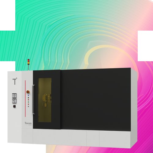
High-Throughput, Multi-Scale Dynamic micro-CT for Imaging in Real-Time Tescan UniTOM® XL, and its smaller version, CoreTOM® are designed for research environments where temporal resolution, imaging flexibility, and experimental throughput matter. A high-power X-ray source, fast large-field detector, and automated multi-sample workflows work together to keep your lab focused on insight, not setup. This results in faster scans, richer data, and true 4D visualization of materials in action. - High temporal resolution for dynamic, in situ, and continuous 3D imaging - Large-sample and multi-scale capability with a spacious scanning envelope - Optimized throughput and reproducibility through automated batch scanning - Seamless workflow control with Acquila™ and Panthera™ software integration WHERE TESCAN UNITOM® XL MAKES THE DIFFERENCE High Temporal Resolution without Compromise: Capture real-time sample changes with continuous 4D scanning at temporal resolutions under three seconds, without sacrificing image quality or stability. Optimized Throughput for Shared Facilities: Maximize productivity through automated batch scanning and rapid acquisition workflows. Tescan UniTOM® XL supports multiple users and high daily sample volumes with consistent results. Flexible Imaging for Every Sample: From centimeter-scale components to fine structural details, the versatile sample stage and ten motorized axes accommodate a broad range of sample sizes and orientations. True Multi-Scale Imaging: Volume of Interest Scanning (VOIS) enables detailed high-resolution imaging within larger samples — providing both context and clarity in a single workflow. In Situ and Dynamic Experiment Capability: Perform uninterrupted insitu studies with slip-ring technology and optional external connection interfaces, supporting mechanical, thermal, or chemical testing during live scanning. Seamless Workflow Integration: Acquila™ and Panthera™ software environments manage acquisition, reconstruction, and visualization — delivering complete experimental control from setup to analysis. Reliable, Non-Destructive Analysis: Generate repeatable, quantitative 3D and 4D datasets without altering the sample, ensuring structural integrity for ongoing or correlative studies. Tescan UniTOM® XL in Materials Science — High-resolution, non-destructive imaging for structural and dynamic analysis - Non-destructive 3D characterization of pores, cracks, inclusions, fibers, interfaces, and internal defects - Multi-scale imaging connects whole-sample context with micron-scale structural detail - In situ and continuous 4D imaging track deformation, cracking, phase changes, and failure mechanisms over time Tescan UniTOM® XL supports material research from routine 3D characterization to advanced time-resolved studies. Researchers can quantify the internal structure of intact materials in 3D, including morphology, porosity, defects, and interfaces, then extend the same workflow into 4D when they need to observe how those structures evolve under load, heat, or chemical exposure. The result is reproducible, non-destructive insight into both material architecture and performance over time. Tescan UniTOM® XL in Semiconductors — High-resolution 3D and 4D micro-CT, from packages to complete electronic assemblies - Non-destructive 3D inspection of complete devices, from small consumer electronics to larger assemblies such as laptops - Large field of view preserves full-device context while enabling analysis of internal components, connections, batteries, PCBs, and housings - High image quality at practical scan times supports failure analysis, design validation, and quality investigation without disassembly - 4D capability can support selected mechanical or thermal reliability studies where internal changes need to be followed under controlled conditions Tescan UniTOM® XL enables researchers and engineers to inspect consumer electronics and electronic assemblies in 3D without opening, cutting, or disassembling the device. Its large field of view makes it possible to image anything from compact devices and individual modules to larger assemblies such as laptops, while preserving the spatial relationship between components. Users can investigate batteries, printed circuit boards, connectors, housings, adhesives, interfaces, voids, cracks, assembly defects, and other internal features that influence reliability and performance. For selected reliability workflows, Tescan UniTOM® XL can also extend from static 3D inspection to 4D imaging, helping teams observe internal changes during controlled mechanical or thermal testing. Tescan UniTOM® XL in Batteries — High-resolution 3D and 4D micro-CT for battery structure, operation, and degradation - Non-destructive 3D analysis of battery cells, electrodes, and solid-state interfaces - Multi-scale imaging captures structure from electrode level to full-cell context - Operando 4D imaging tracks swelling, cracking, electrolyte movement, and degradation during cycling or stress testing Tescan UniTOM® XL supports battery research from static 3D characterization to in-situ 4D experiments, enabling time-resolved micro-CT imaging of batteries under realistic operating conditions. Fast acquisition and continuous rotation support uninterrupted observation during charge–discharge cycling or thermal stress. This enables clearer insight into electrode design, cycle-life behavior, and safety performance. Tescan UniTOM® XL in Life Science — Versatile 3D micro-CT for biological, clinical, forensic, and natural history research - Non-destructive 3D imaging of biological specimens, tissues, and natural history collections - Large field of view supports a wide range of sample sizes while preserving anatomical and morphological context - High image quality at practical scan times for soft tissue, stained samples, mineralized structures, fossils, and dense materials - 4D capability supports biomechanics studies where deformation, motion, or mechanical response needs to be followed over time Tescan UniTOM® XL enables life science researchers to visualize internal structures in 3D across an exceptionally broad range of specimen types and sizes. From delicate biological samples and human tissues to larger developmental, clinical, forensic, and natural history specimens, the system provides the flexiblity needed for multi-user research environments. Its large field of view and strong image quality help preserve whole-sample context while revealing fine internal detail, supporting non-destructive investigation of valuable, rare, or difficult-to-prepare samples. In addition, in situ connectivity and 4D imaging can support time-resolved imaging to connect morphology with mechanical behavior. Tescan UniTOM® XL in Geoscience — High-resolution characterization of geological samples, from 3D pore networks to 4D fluid flow - Non-destructive 3D characterization of pore networks, fractures, grains, mineral phases, and core heterogeneity - Large scanning envelope accommodates full-size rock cores up to 1 m tall - In situ and 4D imaging capture fluid flow, trapping, fracture dynamics, and mineral–fluid interactions over time Tescan UniTOM® XL, and its smaller version, Tescan CoreTOM® support geoscience workflows from static 3D rock characterization to dynamic 4D experiments. Researchers can quantify pore networks, fractures, mineral distributions, and core-scale heterogeneity in 3D, then observe how these structures affect fluid migration, trapping, and deformation under relevant experimental conditions. The systems’ large sample capacity and dynamic imaging performance help connect pore-scale measurements with reservoir-scale models. Tescan UniTOM XL in Materials Science — High-resolution, non-destructive imaging for structural and dynamic analysis - Multi-scale micro-CT enables visualization of structures from millimeters down to microns - Continuous 4D imaging reveals deformation, cracking, or phase changes in real time - Insitu interfaces allow live observation under thermal, mechanical, or chemical stimuli Tescan UniTOM XL supports material research where understanding internal structure and evolution over time is essential. Its combination of high temporal and spatial resolution enables researchers to quantify changes such as fracture propagation, composite failure, and pore evolution. The result is a reproducible, non-destructive insight into material performance and durability. Tescan UniTOM XL in Energy and Battery Research — Dynamic micro-CT for studying material behavior during operation - Real-time visualization of electrode swelling, and electrolyte movement - Non-destructive analysis of battery cells, electrodes, and solid-state interfaces - Multi-scale imaging captures microstructural evolution from particle to full cell TESCAN UniTOM XL enables time-resolved micro-CT imaging of batteries under realistic operating conditions. Fast acquisition and continuous rotation support uninterrupted observation during charge–discharge cycling or thermal stress. This enables clearer insight into electrode design, cycle-life behavior, and safety performance. Tescan UniTOM XL and CoreTOM in Geoscience and Porous Media — In situ observation of fluid flow and structural changes in geological samples - High temporal resolution captures fluid flow, trapping, and fracture dynamics - Large scanning envelope accommodates full size rock cores up to 1m tall - Non-destructive imaging preserves sample integrity for repeated testing Tescan UniTOM XL, and its smaller version, CoreTOM enable geoscientists to observe pore-scale processes in real time. Researchers can follow fluid migration pathways and mineral–fluid interactions in reservoir rocks, extending to CO₂ and H₂ storage scenarios. The system’s large sample capacity and dynamic imaging performance make it ideal for correlating laboratory experiments with reservoir-scale models. TESCAN UniTOM XL in Industrial and Manufacturing Research — High-throughput 3D and 4D imaging for process optimization and quality assurance - Non-destructive inspection of components and assemblies for defect detection - Batch scanning and automated reconstruction for multi-user laboratory environments - Multi-scale imaging supporting analysis from part-level structure to micro-defect formation TESCAN UniTOM XL delivers fast, repeatable imaging that supports process validation and failure analysis in industrial R&D and quality control. Its combination of throughput, resolution, and large-sample handling reduces inspection time while maintaining quantitative accuracy, ensuring reliable evaluation of manufactured materials and components. Tescan Acquila™ and Panthera™ Software Ecosystem — AUTOMATION THAT STREAMLINES 4D IMAGING WORKFLOWS Tescan UniTOM® XL integrates seamlessly with the Acquila™ and Panthera™ software environment, providing coordinated control of acquisition, reconstruction, and visualization. Dynamic CT experiments may run without interruption, with reconstruction and analysis carried out alongside acquisition to maintain workflow efficiency, ensuring uninterrupted operation and maximum throughput. The software suite offers automated workflow management, live monitoring, and real-time visualization in an intuitive interface that reduces setup time and ensures reproducible results. For dynamic or in situ studies, Acquila™ and Panthera™ synchronize imaging with external rigs and detectors to maintain full experimental control throughout the 4D imaging process.
CATEGORY
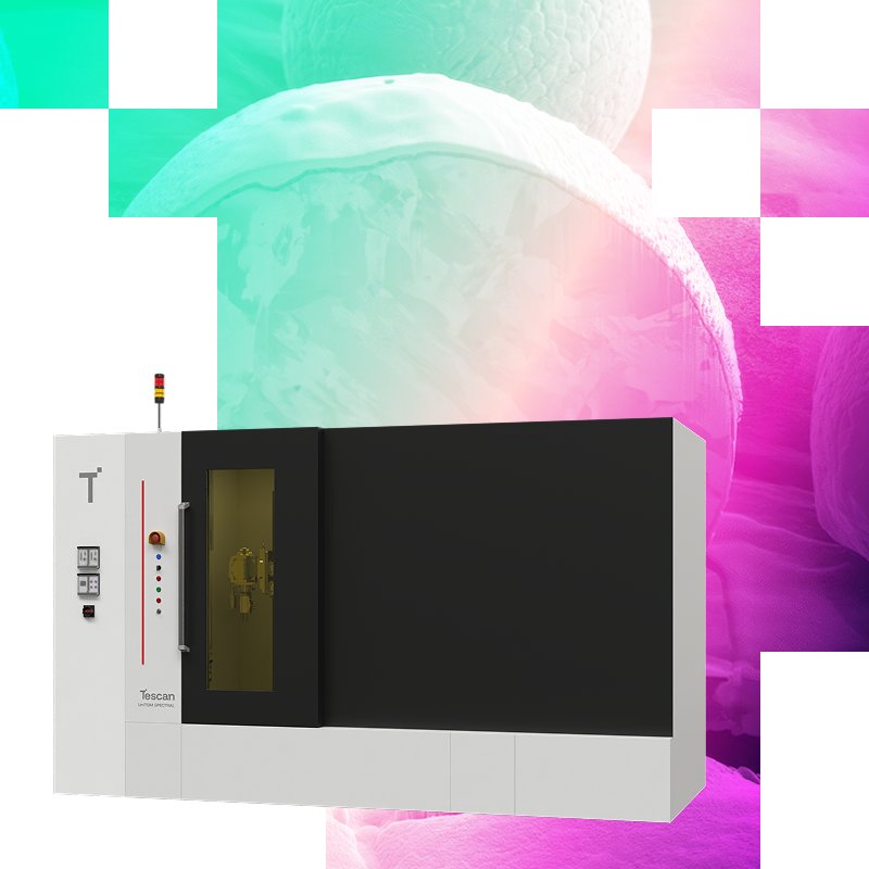
Non-destructive elemental information from any point inside a sample Tescan Spectral CT is designed for laboratories that need non-destructive 3D elemental insight alongside structural imaging. As an add-on to the Tescan UniTOM® XL, it uses full-spectrum, multi-energy acquisition with a dedicated secondary spectral detector to capture elemental signatures across the sample. With Tescan Spectral Suite™ and Acquila™ software working together, users can target regions of interest, reconstruct spectra, and correlate elemental with structural information in one workflow. This delivers clear material contrast and reliable elemental identification in every scan. - Non-destructive spectral micro-CT for 3D elemental information at any point inside the sample - TrueContrast™ multi-energy imaging to differentiate similar-density materials conventional CT cannot - Material identification using k-edge detection for absolute elemental identification without prior knowledge - Material identification using effective atomic number (Z-eff) and density extraction - Spectral Suite™+ Acquila™ integration for VOI targeting, spectral reconstruction, and analysis Full-Spectrum Imaging: Capture the entire X-ray spectrum during every scan to reveal both structural and elemental information in one dataset—no additional instruments or destructive preparation required. True Multi-Energy Contrast: Differentiate polymers, metals, or biological tissues with TrueContrast™ multi-energy imaging, delivering clarity where conventional micro-CT reaches its limits. K-Edge Element Detection: Identify and map precious metals and elements with absolute certainty using K-edge detection, enabling confident analysis of complex or unknown materials. Non-Destructive compositional Insight: Visualize internal composition and extract material density without cutting or altering samples, preserving integrity for further testing or archival studies. Integrated Spectral Suite™: Control acquisition, reconstruction, and spectral analysis through Spectral Suite™ software, fully synchronized with Acquila™ micro-CT control for streamlined workflows. Versatile Multi-Disciplinary Applications: From mineral cores to polymer composites and biological specimens, SPECTRAL CT expands UniTOM® XL performance into new domains of 3D elemental and structural research.
CATEGORY
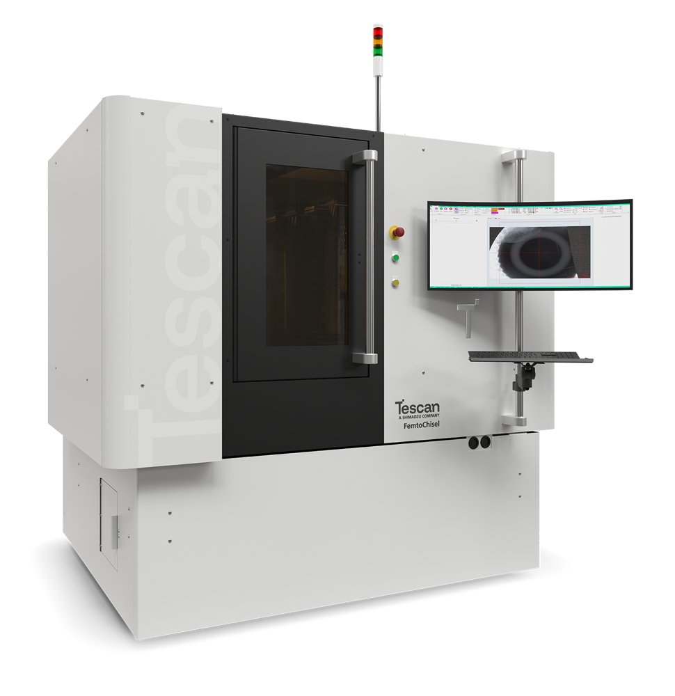
Femtosecond Laser for High-Precision Semiconductor Sample Prep Tescan FemtoChisel™gives you nanometer-level precision and high-throughput intelligent laser processing for semiconductor research and failure analysis. It delivers pristine surfaces while significantly reducing the need for subsequent FIB polishing. Accelerate turnaround in device analysis and failure investigation for current and next-generation semiconductor materials. Achieve Pristine Laser Surfaces for Faster Semiconductor Analysis Tescan FemtoChisel™ is a femtosecond laser micro and nano machining system for sample preparation, micro and nano fabrication, failure analysis, and surface modification. Designed for semiconductor, glass processing, materials research, and biomedical device manufacturing workflows, it helps users access buried regions of interest, prepare clean surfaces, and accelerate analysis. Key benefits - Intelligent Multi-Gas Processing - Laser Protective Layer - Multi degrees of freedom stage with tilt and wafer-level rotation - Correlative Machine Vision & Targeting - Software-Selectable Laser Wavelength - Real-time surface and ablation monitoring with material adaptability
CATEGORY
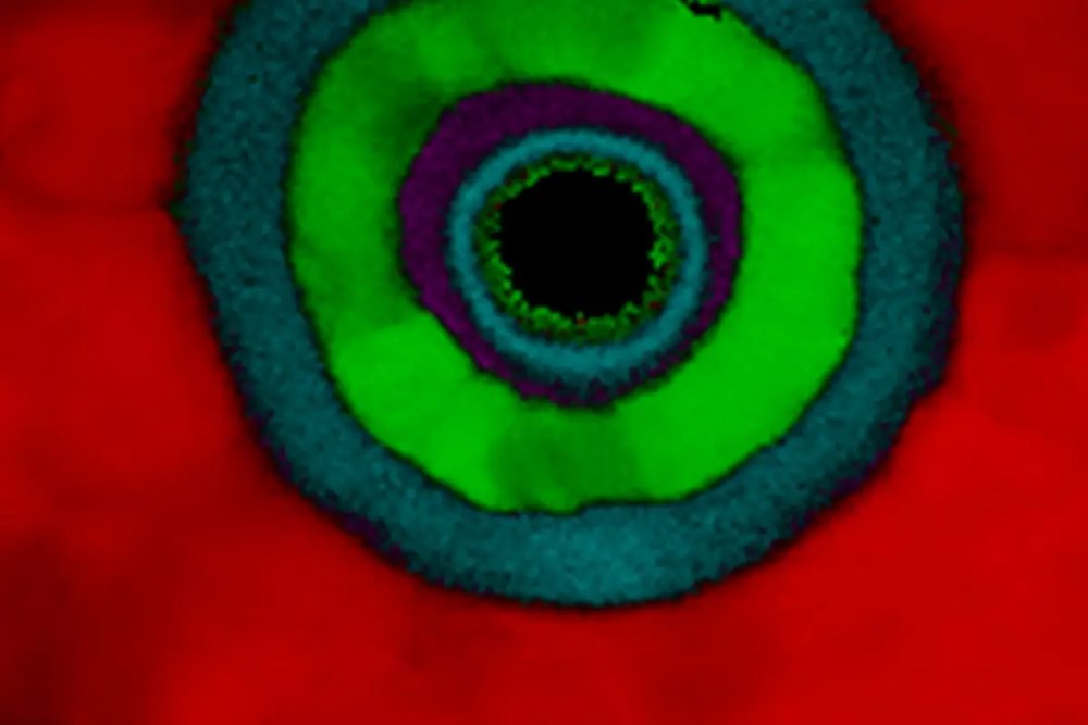
Analytical STEM with integrated beam precession for enhanced 4D-STEM and 3D-ED structural studies Tescan TENSOR™enables fast and intuitive sample navigation and identification of regions of interest with their st Discover Tescan’s Nanoscale Insights with Tescan TENSOR™ Analytical STEM Assess the morphology, composition, and structure of analyzed samples with clarity, speed, and confidence to gain insights into their physical properties and function. Tescan’s analytical STEM solutions provide the necessary tools needed for advanced materials research, semiconductor analysis, and detailed nanoscale characterization, down to the atomic level. With fully integrated precession-assisted diffraction, and synchronized 4D-STEM and EDS workflows, you get immediately meaningful results and actionable insights with practical usability—whether you need to assess sample crystallinity, identify the distribution of different phases, validate strain engineering, or support process development and failure analysis.
CATEGORY
