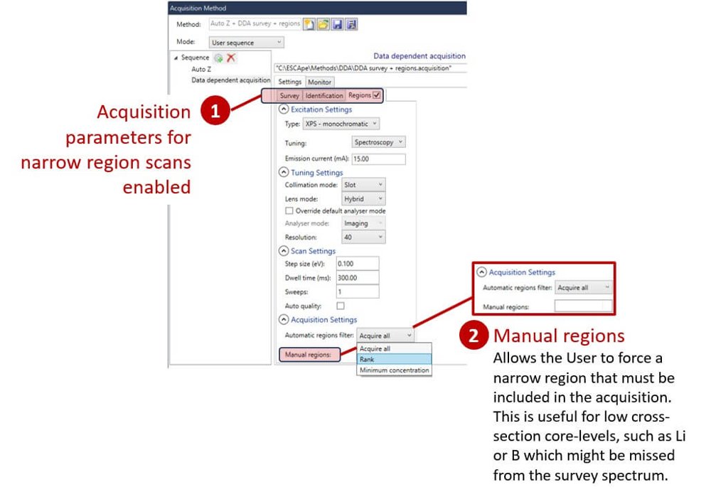
ESCApe software has been developed in close collaboration with our Users. We understand that it is the interface between the User and spectrometer. Removing Users’ pain-points from the workflow ensures that using a modern AXIS spectrometer is as simple as possible. Incorporating acquisition and processing capabilities within the same software blurs the distinction between the two functionalities. As we move into the age of artificial intelligence, ESCApe software is well placed to allow acquired data to inform further acquisition steps. Complete instrument control Whilst ease of use and simplicity are extremely important, we understand that there are times when complete, manual control of the spectrometer is a requirement. ESCApe software can be configured with hierarchical User access. With full access, manual mode provides access to direct control, instrument tuning and instrument configuration environments. There is also comprehensive component parameter logging allowing interrogation of key operating parameters instantaneously and over extended periods. This capability also provides valuable insight into preventative maintenance intervention and allows remote engineer support and fault finding. In acquisition queue mode, the instrument status window provides clear, visual confirmation that the instrument is operational. Similarly a vacuum mimic diagram reports real-time status of vacuum components, including values and pumps. Automated acquisition A strength of the ESCApe software is the level of automation. Confidence in unattended acquisition means that the spectrometer can perform extended analysis across numerous samples. Ambiguity of elemental identification has been removed by development of a robust peak-ID algorithm. Peak-ID also extends to spectra generated in HAXPES mode, using the Ag L α excitation source. ESCApe takes automated sample analysis to a new level through Data Dependent Acquisition (DDA). This automation incorporates data processing into experimental methods freeing the analyst from the more routine aspects of data acquisition. Sample analysis height is optimised from the position defined on the optical microscope images prior to acquisition of a survey spectrum acquired in highest sensitivity mode. Subsequently the survey spectrum is automatically processed to identify the elements present and their concentrations. This information is then used to determine the regions for high resolution analysis and subsequent chemical state determination. The quantification routine includes a measure of the error in the atomic concentration. High resolution regions can be acquired to a specified signal to noise ratio. Using DDA you can be confident of producing exceptional quality data. Data dependent acquisition (DDA) The standard data acquisition workflow for AXIS using ESCApe software uses pre-defined standard methods. Using these methods ensures that the instrument parameters are correctly defined, whilst also allowing the expert User to tailor the acquisition as necessary. Automated data acquisition can be taken a step further using the newly developed DDA. Following an ‘auto-Z’ acquisition which puts that sample at the correct analysis height, a large-area survey spectrum is acquired. The DDA will run an auto-peak ID process to identify and quantify the elements present in the survey spectrum, followed by the acquisition of narrow region spectra at high resolution for the primary peaks of the elements identified. As you would expect from intelligent software, there are a number of different triggers defining which narrow regions are acquired. These include: Acquire all– acquires region scans of all identified primary core levels. Rank – used to define a specified number of most abundant regions acquired based on their atomic concentration. Minimum concentration – used to define regions acquired based on a set minimum atomic concentration. There is also a Manual regions option, which allows the User to force a narrow region that must be included in the acquisition. This is useful for low cross-section core levels, such as Li or B which might be missed from the survey spectrum. Group array analysis Array analysis is an exciting way of acquiring and visualising large dataset information in a correlative way. With some samples, elemental or chemical distribution may not be homogeneous across the surface. Differences may exist over extended areas of the sample. By submitting an array analysis data can be acquired from discrete positions across the sample. Spectra are quantified and the information displayed graphically as an overlay on the optical image. This simple approach to large dataset acquisition and display allows easy identification of regions of interest or correlation between elemental/chemical distribution across the sample. Supporting additional analytical techniques Additional analytical techniques complement the surface chemical information provided by XPS. ESCApe software supports acquisition and data processing of most additional techniques available for the AXIS spectrometers. Using optional excitation sources, such as the HAXPES high energy Ag L α X-rays or low energy UV He discharge, is as simple as selecting the appropriate acquisition method. Seamless integration of these additional techniques ensures that they can be easily used, removing the barriers of complexity often associated with secondary analytical techniques. Auger electron spectroscopy, scanning Auger microscopy and scanning electron microscopy are supported by ESCApe. Coincidence of the XPS and Auger analysis extends the useful information that can be derived from a sample. The significantly smaller probe size provided by Auger analysis allows surface analysis of regions of interest too small to analyse with XPS. ESCApe software also integrates a number of data processing capabilities required for data reduction specific to optional techniques. ESCApe data processing ESCApe software provides a comprehensive toolkit for data processing. We’ve automated peak-ID and survey spectrum quantification. Powerful data reduction routines allow the User to fit complex models to spectral envelopes. Data reduction can be simplified by dropping a saved model onto an unprocessed spectrum. A linear least squares fit is applied to optimise the fit of the model to the acquired data. Extraction of depth distribution information from angle-resolved XPS analysis is achieved using Kratos developed maximum entropy method software embedded within ESCApe. ESCApe data processing extends to imaging data, with easy generation of quantitative XPS images. Post-acquisition spectral generation from spectromicroscopy data is seamlessly displayed, providing an innovative approach to determining lateral distribution of surface chemistry. Result reporting Once data reduction is complete, it’s important to be able to distribute the results. ESCApe software offers a number of useful output options. An Excel add-in allows experimental datasets to be opened with the same tree structure used within ESCApe software. A complete description of the spectrum, curve fits, envelope background and transmission function are available within Excel. Batch output for multiple spectra is possible. Data is also transferable using the VAMAS format, making it compatible with CasaXPS.
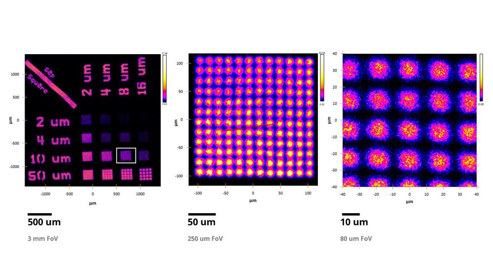
Materials scientists and engineers needing quantitative surface chemistry from the top ~10 nm of a sample using and instrument with high sample throughput and automation. The instrument is used in both academic research and industrial labs. The AXIS Supra+ is set apart from any other spectrometer by complete automation through computer control of sample handling and instrument parameters. Unattended sample holder transfer and exchange during analysis is achieved through coordination of the Flexi-lock sample magazine and sample analysis chamber auto-stage. ESCApe integrated acquisition and processing software allows the AXIS Supra+ to perform to its maximum capability and provides an easy interface with the spectrometer. Large area, high sensitivity XPS: The AXIS Supra+ is optimised for chemical state X-ray photoelectron spectroscopy. Efficient collection of photoelectrons combined with high transmission electron optics ensures unrivalled sensitivity and resolution at large analysis areas. As well as conventional scanned acquisition, spectra may be acquired in fast, unscanned snap-shot mode in less than a second, making use of the 128-channel Delay-Line Detector (DLD). Key attributes include: - Easy detection of light elements. - Excellent signal-to-noise, even at low concentrations. - Fast data acquisition. - Scanned or snap-shot spectral acquisition modes. Highest energy resolution: The fundamental requirement of any spectrometer is the best possible energy resolution. High energy resolution, where the spectrometer does not contribute to the broadening of the photoemission peaks, is critical for the accurate measurement of small chemical shifts. The AXIS Supra+ has a large 500 mm Rowland circle monochromated Al Kα X-ray source and optimised electron optics, ensuring excellent chemical resolution. Advantages of high spectral energy resolution include: - Unambiguous identification of chemical shifts. - Guaranteed energy resolution on insulating and conducting samples. Fast parallel XPS imaging: The lateral distribution of elements or chemistry at the surface is measured by XPS imaging. The AXIS Supra+ acquires fast, high spatial resolution parallel images. Parallel image acquisition has the advantage that it is significantly faster and achieves higher spatial resolution than the more conventional rastered beam approach. Parallel imaging may also be combined with stage movements to acquire a ‘stitched’ image, capable of generating images over several millimetres with a spatial resolution of several microns. Capabilities provided by parallel imaging include: - Ultimate spatial resolution of 1 micron at the highest magnification. - High energy resolution, chemical state imaging. - Quantitative imaging – the unique spherical mirror analyser and delay-line detector can provide quantitative chemical state images. - Spectromicroscopy – easy acquisition of spectra from image datasets providing a spectrum at each pixel. Small spot, selected area spectroscopy: The AXIS Supra+ small spot performance guarantees high sensitivity in small spot mode by optimising X-ray illumination of the sample with the analysis area selected. The standard, high flux monochromatic X-ray source is ideal for analysis that requires extremely high sensitivity performance from small selected areas. Key selected area spectroscopy attributes include: - Pre-defined small spot analysis areas. - Optimised X-ray illumination for improved selected area performance. - Automated aperture and iris in the electrostatic lens column to form a virtual probe at the sample surface. - Selected area spectra can be acquired using either the monochromatic or achromatic X-ray sources. Unrivalled instrument automation: The AXIS Supra+ has complete automation of sample handling which sets it apart from any other spectrometer. Auto sample exchange at the end of an experiment allows continuous operation. Unprecedented levels of automation extend to routine maintenance aspects such as computer controlled bake-out and subsequent degassing of filaments. Automation also includes the X-ray source and monochromator mirror so that calibration and switching between Al K α and optional Ag L α excitation sources is completely computer controlled. The motorised multi-position anode ensures continued optimised performance. Intuitive ESCApe software combined with instrument automation ensures that the spectrometer is easy to use, even for less experienced operators. More complex data acquisition can be undertaken with confidence that the instrument will operate reproducibly. Multi-technique & sample modification: Optional excitation sources include an achromatic Al/Mg X-ray source, Helium discharge lamp for ultraviolet photoemission spectroscopy (UPS) and a high energy Ag L α monochromated X-ray source for Hard X-ray photoelectron spectroscopy (HAXPES). The ability to probe unoccupied electronic orbitals is provided by addition of Inverse Photoemission Electron Spectroscopy (IPES). Addition of a field-emission electron source adds Auger electron spectroscopy (AES), scanning Auger mapping (SAM) and secondary electron microscopy (SEM) capability to the instrument, with the advantage of greater lateral resolution relative to XPS. These additional techniques are coincident with the XPS analysis position giving complementary insight to the sample. Importantly, addition of these techniques does not compromise the market leading XPS performance. Sample preparation and surface modification options can be accommodated on the introduction chamber, also known as the flexilock. These options include sample heat and cool, air sensitive sample transporter, broad spot ion source, crystal clever and glove box. We have designed a number of optional sample holders allowing in-situ and in-operando measurements to be achieved. Sample holders allow electrical charge-discharge cycling, biasing with/without additional heating. A third chamber can be configured to provide a dedicated UHV environment for surface science studies. Typical configuration equips this chamber with a manual stage and optional characterisation techniques including low energy electron diffraction (LEED), inverse photoemission spectroscopy (IPES), quadrupole secondary ion mass spectrometry (SIMS). A gas reaction cell, useful for high temperature and pressure sample modification, is available as an accessory. Many of the optional items listed above are available as upgrades post-installation. We also highlight that Kratos Physicists and Mechanical Engineers are able to provide solutions for bespoke sample handling solutions and additional analytical technique integration for the AXIS Supra+. Hard X-ray photoelectron spectroscopy (HAXPES): There are advantages to using higher energy X-ray sources to acquire photoelectron spectra. The AXIS Supra+ uses monochromated Ag Lα with photon energy of 2984.3 eV. Changing between the Al K α and Ag L α excitation source is fully computer controlled. The analysis position for the 2 sources is set up to be coincident. Quantification using the HAXPES source is calculated in an analogous way to the standard Al excitation, where the spectra are corrected for spectrometer transmission and core level relative sensitivity factor. These include: - Access to higher BE core levels. - Increased sampling depth (relative to Al Kα). - Shifting Auger transition overlaps with XPS core levels. Our introductory video gives an overview of the capabilities of the AXIS Supra+. See data acquisition and sample holder exchange in action. Learn about some of the many applications of the AXIS Supra+. Electron analysers: The AXIS Supra+ combines a 165 mm mean radius hemispherical sector analyser (HSA) for spectroscopy mode with a patented spherical mirror analyser (SMA). This unique combination allows high energy and spatial resolution in both spectroscopic and imaging modes. AXIS technology for electron transfer from the sample to the analysers includes our patented magnetic immersion lens and electrostatic lens column. The patented electron only charge neutraliser is incorporated at the end of the lens column, directly above the sample. Delay-line detector: The delay-line detector (DLD) is used for counting photoelectrons in both spectroscopy and imaging modes. It comprises a multi-channel plate (MCP) stack above a 2-dimensional delay-line winding, a pre-amp and fast counting electronics. The DLD has three operating modes; conventional scanned spectroscopy mode, unscanned snapshot spectroscopy and 2D, parallel imaging mode. Auto-stage: Automated 5-axis precision manipulator. The stage is fully integrated with the acquisition software, allowing unattended sample exchange. The sample stage can be equipped with the optional heat and cool accessory. Data system: ESCApe acquisition and processing software is developed and maintained by Kratos’ software group. The software runs on Microsoft Windows and makes User interaction with the spectrometer as simple as possible. As we move towards data dependent acquisition, automated peak identification and subsequent narrow region acquisition and reporting has been realised. Electronics: The instrument control units are accommodated underneath the work desk. Discrete units are used to control the numerous components that comprise the instrument. Additional accessory control units are housed within the same enclosure. The instrument has a relatively compact footprint. Ion sources: The AXIS Supra+ can be specified with a standard monatomic Ar+ source (Minibeam 4) or optional Gas Cluster Ion Sources (GCIS, Minibeam 6 or VCIS). Both types of ion sources are fully integrated into the ESCApe software for ease of use, including automated gas handling. The ion sources are all capable of operating in He+ ion mode for optional Ion Scattering Spectroscopy (ISS).
CATEGORY
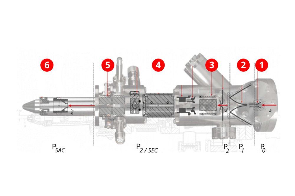
When introduced almost a decade ago, the Minibeam 6 gas cluster ion source represented a step change in sputter depth profiling capability. It provided a clean, reliable way to successfully sputter depth profile through polymer materials. This capability was quickly recognised and the use of cluster ions for depth profiling increased almost exponentially. For inorganic materials, the sputter yields when using cluster ions are significantly lower. This means that depth profiling experiments are much longer than organic depth profiles. The lower etch rates can be mitigated somewhat by using smaller, higher energy cluster ions, with the major advantage of less chemical damage when compared to conventional monoatomic sputtering. Gas cluster operating mode: Large Ar clusters are formed by isentropic adiabatic cooling of the gas as it expands from high pressure (P 0) through a nozzle into the vacuum of the source region (P 1/ P 2). The Ar n clusters are ionised by electron impact, extracted from the source region and accelerated along the ion column where a Wien velocity filter is used to select a median cluster size between 500 and 3000. These cluster ions may be accelerated up to a maximum of 20 keV such that the energy per atom, or partition energy, can be varied from < 2 eV (5 keV, Ar 3000+) to 40 eV (20 keV, Ar 500+). A subsequent ion channelling bend in the focusing column removes energetic neutrals. The beam is then focused to a minimum spot size of 250 μm on the sample. Scan plates allow the beam to be rastered across the sample surface to create an even depth profile. The ability to generate Ar n+ions with different cluster sizes and acceleration energies means successful depth profiles can be generated from the greatest range of materials. Large clusters, where n 2000, are useful for depth profiling organic layer systems whilst smaller clusters of n=500 accelerated to high energies have been shown to be necessary in retaining chemistry of inorganic materials during depth profiling and sample cleaning. Monatomic operating mode: The ion source can be operated in monatomic Ar+ ion mode for conventional depth profiling inorganic and metallic samples. The sputter yield for this class of materials is known to be much higher for monatomic than cluster ions making Ar+monatomic mode important for depth profiling in realistic experimental times. Ion acceleration voltage can be varied between 500 eV and 5 keV. Hardware & software: The standard GCIS configuration provides differential pumping of the ion source by two dedicated turbo molecular pumps with additional pumping provided by the sample entry chamber. An automated gas handling manifold is provided to supply both high and low pressure for cluster and monatomic modes respectively. The GCIS control unit is computer controlled through the ESCApe software allowing unattended operation of and switching between Ar n+ cluster and Ar+ or He+monatomic modes. For routine operation the User interface allows simple recall of standard operating methods for sample cleaning and depth profiling in cluster and monatomic modes. Operating parameters of the ion source are continuously displayed allowing key parameters to be monitored along with a vacuum mimic diagram showing the status of valves and pumps. For multilayer sputter depth profiling, sample rotation is known to reduce ion induced sample roughening and maintain interface depth resolution. Compucentric rotation about the analysis position is supported when the sample is mounted on the Z-rotation sample holder The multimode gas cluster ion source provides the capability to depth profile polymer, inorganic and metallic samples through a choice of giant gas cluster or monatomic argon ions. Easy control of both the cluster size and ion energy allows the appropriate partition energy (energy per atom) to be used for the application whilst maintaining suitable sputter yields per incident ion. Small clusters at high energy (20 keV, Ar 1000+) are useful for depth profiling inorganic materials and demonstrate significantly improved retention of stoichiometry when compared to monatomic Ar+ profiles. Large, lower energy clusters (5 keV, Ar 3000+) are employed for removal of surface contamination of polymer materials whilst medium energy, large clusters (10 keV, Ar 3000+) are suitable for extremely deep profiling of organic materials. The GCIS provides significant enhancement of capabilities over the standard monatomic ion source. The use of large, low energy cluster ions ensures retention of chemistry when profiling soft, organic materials. Alternatively, selecting a small cluster, high energy mode facilitates the sputtering of inorganic materials, whilst minimising preferential sputtering and chemical damage. Using the monatomic mode means that metallic thin films can also be sputtered.
CATEGORY
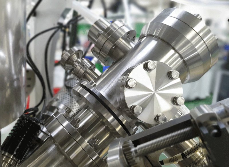
Monatomic Ar+ Ion Source. This ion source is designed to produce a stable, high current, monatomic Ar+ ion beam with energies from 0.5 to 4 keV. High ion flux guarantees excellent sputter depth profiling etch rates. The Minibeam 4 is ideally suited to sputter cleaning and depth profiling inorganic and metallic films.It may also be used to generate low-energy He+ ions for use with ion scattering spectroscopy (ISS). The Minibeam 4 is an ideal sputter source for sample cleaning and sputter depth profiling inorganic or metallic thin-films and multilayer materials. It is capable of generating a high flux, focussed beam of monatomic Ar+ ions. The ion source is fully integrated with the ESCApe data-system, which automates gas handling. Monatomic operating mode: The ion source is operated in monatomic Ar+ ion mode for conventional depth profiling inorganic and metallic samples.Ion acceleration voltage can be varied between 500 eV and 4 keV. Floating ion column: The ion column is electrically isolated from the ion source body which allows the source to be operated in ‘floating’ mode. In this mode of operation, ions generated at low potential are accelerated to high energies for efficient transmission through the ion column. This focused, high current, beam is then retarded back to low energy, typically 500 eV, before leaving the ion source. The resultant high flux, low energy ion beam is used for better retention of layer interface during profiles. Studies demonstrate that lower beam energies cause less interface mixing and sample roughening at the bottom of the sputter crater. Minibeam 4 architecture: The ion source comprises of an electron impact ionisation region fitted with 2 filaments. The ion extraction current forms a negative feedback loop with the filament current thus allowing automatic stabilisation of the ion source current. In floating mode the ions are extracted and accelerated into the floating ion column. Following beam formation an ion channelling bend in the focusing column eliminates neutral species. An objective lens focuses the ion beam to a minimum spot size of 600 um and scan-plates raster the beam across the sample surface. Differential pumping is provided by the turbo molecular ion pump of the Flexi-lock. Software control: Our Minibeam 4 is fully integrated with ESCApe data system. Sample cleaning and sputter depth profiles are defined as standard acquisition methods for easy, routine use of the ions source. Operating parameters are defined in look-up tables so that reproducible etch rates are achieved. When required for creation of additional tuning modes or detailed observation of operating conditions, readbacks for critical operating parameters are constantly available. These values can optionally be written to log files. As expected from a fully automated instrument, the ion source can be used to generate unattended sputter depth profiles including full control of Ar (gas). Compucentric rotation: If samples are mounted on the appropriate rotation sample holder, compucentric rotation can be simply achieved. The sample is rotated about the analysis position in 90 o steps for each sputter cycle. Usefully, the sputter position does not need to coincide with the center of rotation of the sample holder, as the ESCApe software will rotate the sample, ensuring the area under investigation remains at the instrument analysis position. This step-wise method, as opposed to continuous rotation, has been shown to result in a more uniform ion fluence within the sputter area for a rastered beam. This in turn will produce better interface resolution between thin layers due to less sputter induced sample roughening. As standard the AXIS Supra+is configured with the Minibeam 4 monatomic Ar+ source. This high flux ion source is fully software controlled and capable of generating depth profiles from a wide range of materials.
CATEGORY
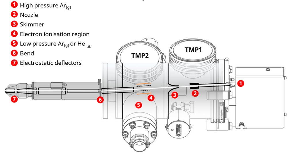
The Versatile Cluster Ion Source enables high-resolution depth profiling of thin films, multilayer structures, and surface coatings. It provides quantitative insights into layer thicknesses, interfacial chemistry, and diffusion phenomena, making it invaluable for materials research, semiconductor fabrication, and corrosion analysis. The technique's ability to detect subtle changes in oxidation states and bonding environments enhances its utility in evaluating complex material systems. The capabilities of the Versatile Cluster Ion Source are outlined below. Cluster ion sample cleaning XPS analysis of exfoliated MoS2 showed carbon and oxygen surface contamination in the survey spectrum (blue). Arn+ cluster cleaning of the sample is shown to remove this surface contamination (red) without change in the high-resolution narrow region scans of Mo 3d / S 2s and S 2p regions. Importantly, the X-ray excited valence band spectra also show no significant change in density of states. Cluster ion depth profiling Depth profiling of polymers typically utilizes Arn+ cluster ions at energies ranging from 5 to 10 keV, which facilitates high sputter rates while preserving interfacial resolution. Minimizing chemical degradation during ion bombardment is essential to maintain the integrity of the material's chemical structure throughout the profiling process. This example presents a 10 keV Arn+ cluster ion sputter depth profile performed on a polymer deca-layer structure composed of alternating 100 nm films of nitrogen-functionalized polyester and methacrylate. The profiling demonstrates consistent interface resolution across all layers, indicating minimal intermixing or degradation during sputtering. Preservation of polymer stoichiometry is confirmed by high-resolution C 1s spectra obtained at etch cycles 2 and 73, corresponding to the first and ninth polymer layers, respectively, validating the chemical integrity throughout the depth profile. Monatomic Ar+ depth profiling A critical objective in sputter depth profiling is the accurate identification and quantification of film thickness. In this example, monatomic Ar⁺ ions were used to profile a mixed metallic/inorganic multilayer structure containing a 20 nm titanium delta-layer. Notably, the Ti layer exhibited partial oxidation throughout its depth, likely resulting from titanium's high reactivity and its tendency to getter ambient oxygen during deposition. The profiling was conducted using the floating column mode, where ions were generated and initially accelerated to 2 keV. The ions were subsequently retarded to 500 eV before exiting the ion source and interacting with the sample surface. This configuration delivers a high ion flux and efficient etch rates while mitigating surface roughening and interlayer mixing typically associated with higher energy beams. It is highlighted that the measured interface resolution between the SiO₂ and Si layers was determined to be 5.5 nm. Dual mode monatomic/cluster profiling To further validate the performance of the VCIS an 85 nm thick poly(lactic-co-glycolic acid) (PLGA) organic capping layer was spin-coated onto a multilayer substrate comprising Au/Ti/SiO₂/Si. Depth profiling was conducted in unattended mode, with 10 etch cycles using 10 keV Arn+ cluster ions, followed by an automated transition to monatomic Ar+ ion etching. Reconstruction of the depth profile used calibrated etch rates specific to each ion species. It is important to note that the apparent thickness of the Au layer is exaggerated due to the minimal sputtering efficiency of cluster ions on metallic films. The use of Arn+ clusters for profiling through the PLGA layer offers the significant advantage of preserving the chemical integrity of the polymer, enabling accurate characterization of its composition and interface structure. Ease of use The VCIS ion source is completely computer controlled, including fully automated gas handling. For depth profiling in either monatomic or cluster modes, the User simply selects the required operating method in the ESCApe acquisition software. Critical operating parameters are available as readbacks and can be logged during operation to ensure repeatability and reproducibility of depth profiles. A vacuum mimic diagram shows the status of valves, pumps and pressures. Compucentric rotation If samples are mounted on the appropriate rotation sample holder, compucentric rotation can be simply achieved. The sample is rotated about the analysis position in 90 o steps for each sputter cycle. Usefully, the sputter position does not need to coincide with the center of rotation of the sample holder, as the ESCApe software will rotate the sample, ensuring the area under investigation remains at the instrument analysis position. This step-wise method, as opposed to continuous rotation, has been shown [1] to result in a more uniform ion fluence within the sputter area for a rastered beam. This in turn will produce better interface resolution between thin layers due to less sputter induced sample roughening. Components of the VCIS 1 High pressure Ar(g) High-pressure gas is essential for the operation of an argon (Ar) cluster ion source due to the physical requirements of cluster formation. To generate Ar clusters (Arn+ where n can range from 10's to 1000's), individual Ar atoms must condense into clusters. This requires a supersaturated vapor phase, which is achieved by expanding high-pressure argon gas through a nozzle into a vacuum. 2 Nozzle The nozzle in a gas cluster ion source plays a critical role in the formation of cluster ions by enabling the supersonic expansion of high-pressure gas into a vacuum. Here's how it functions: - Supersonic Expansion - Cluster Nucleation - Beam Collimation - Flow Regulation The nozzle transforms high-pressure monatomic gas into a stream of condensed clusters suitable for low-damage sputter depth profiling, especially in sensitive materials like polymers and organics. 3 Skimmer The skimmer in a gas cluster ion source serves a crucial role in shaping and stabilizing the supersonic gas jet used for cluster formation. It is a precision component that enhances cluster beam quality by isolating and directing the optimal portion of the supersonic jet, enabling efficient ionization and consistent sputtering performance. 4 Electron impact ionisation region Once clusters are formed, they are typically ionized via electron impact. A dense neutral cluster population—enabled by high gas pressure—improves ionization yield. 5 Low pressure Ar(g) or He(g) For operation in monatomic Ar+ion mode, the Ar gas is leaked directly into the ionization chamber, bypassing the nozzle and skimmer used for cluster generation. The Ar gas pressure is regulated by an auto-regulated leak value. The Ar+ ions are likewise created by electron impact. The Ar gas supplied can be swapped to He, allowing the creation of He+ ions where the Ion Scattering Spectroscopy (ISS) accessory has been supplied. 6 Bend A bend is included in the ion column to eliminate neutral atoms and molecules that are not ionized. Since neutral species are unaffected by electric or magnetic fields, introducing a bend allows only charged ions to follow the curved path, effectively filtering out neutrals and improving beam purity. 7 Electrostatic deflectors Raster plates in an argon (Ar) ion source are used to deflect and scan the ion beam across the sample surface in a controlled pattern. Their primary function is to enable beam rastering, which distributes the ion dose uniformly over a defined area, improving sputter uniformity and minimizing localized damage.
CATEGORY
