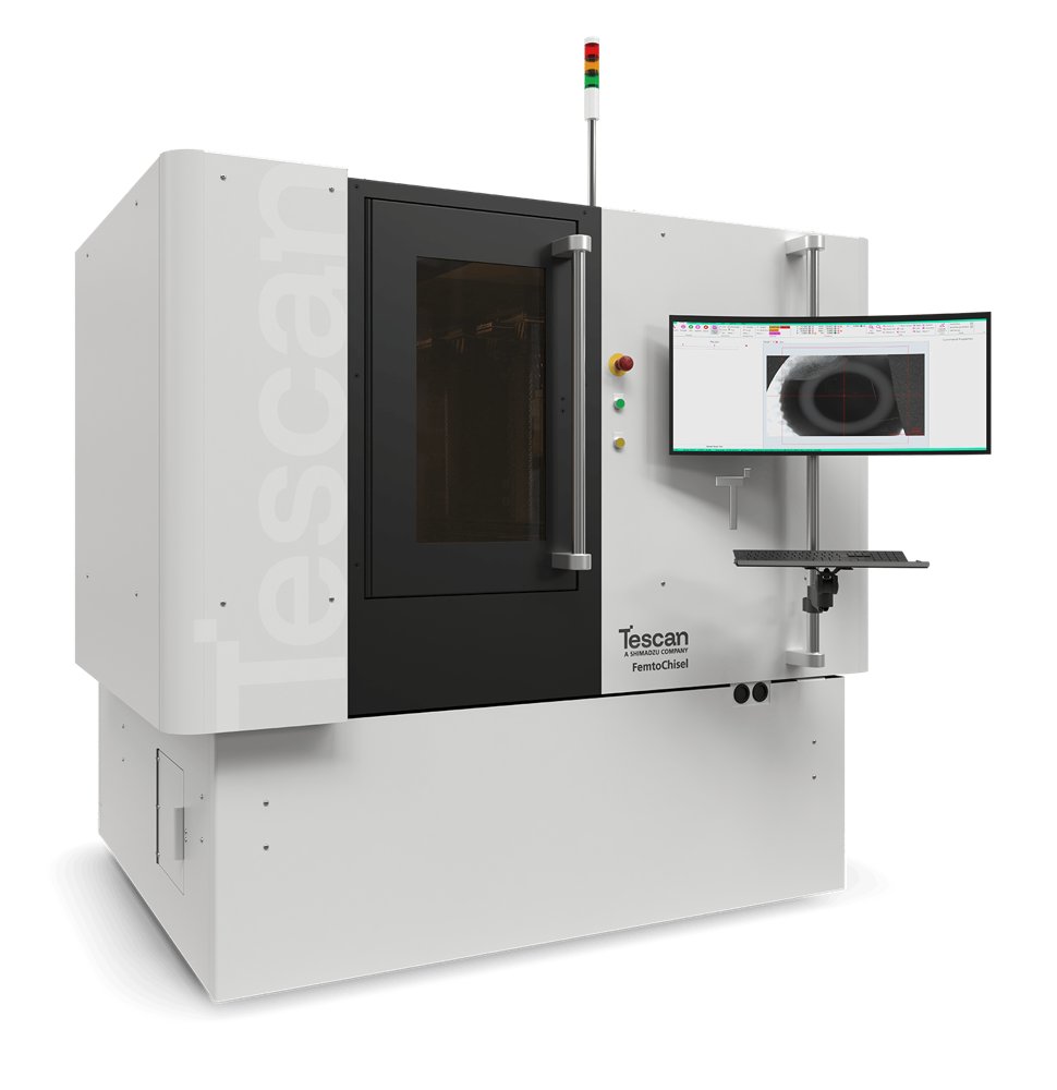
FemtoChisel™ for High-Precision Semiconductor Sample Prep FemtoChisel™ is the next-generation femtosecond laser solution built specifically for semiconductor sample preparation and failure analysis. For too long, the industry has had to compromise between throughput, precision, and Surface Quality. Now all are delivered on one integrated platform.
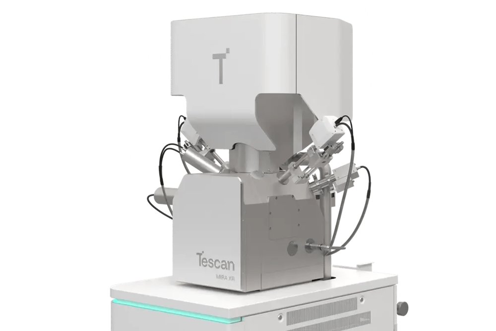
Maximum information from every scan and any sample at the nanoscale Tescan CLARA ™ (UHR-SEM) Ultra-High-Resolution SEM That Reveals More Information from Every Scan Extract more information from challenging materials at low landing energies. CLARA™ combines field‑free ultra‑high‑resolution SEM imaging, advanced secondary electron (SE) and backscattered electron (BSE) detection, and guided automation to help you move from sample navigation to confident insight faster. Designed for nanoscale characterization across diverse materials and workflows, CLARA™ enables you to reveal fine surface structures, material differences, and near-surface features from a single acquisition while maintaining the flexibility required for advanced analytical techniques. CLARA™ gives researchers the resolution, contrast control, and guided operation needed to explore surface detail, material differences, and near‑surface features with confidence. Built on Tescan's BrightBeam™ field-free SEM column technology and operated through Tescan's Essence™ software, CLARA™ supports efficient work across diverse samples, shared instruments, and mixed experience levels. It helps you capture meaningful information with less compromise, less setup, and fewer repeated measurements. - True ultra-high-resolution field-free imaging at low keV Delivers stable nanoscale detail across different samples, including charging, sensitive, and magnetic samples. - Simultaneous UHR SE and BSE imaging with advanced contrast control Combines parallel acquisition of SE and BSE signals with energy-filtered and angle-selective BSE contrast for deeper material insight in a single scan. - Effortless switching between imaging conditions and seamless navigation Powered by In-Flight Beam Tracing® automation for rapid beam optimization, combined with Wide Field Optics® for seamless navigation from macro to nano. - Advanced analytical workflows with open platform flexibility Supports integration of multiple analytical modalities (EDS, EBSD, Raman, etc.) and customizable workflows via custom automation and Python-based scripting. - MultiVac low-vacuum imaging with water vapor and easy switching Improves signal quality and enables stable imaging and analysis of charging and beam-sensitive samples with an uncompromised view field. Where Tescan CLARA™Makes the Difference Reveal More Information from Every Scan Modern nanoscale characterization requires more than resolution alone. CLARA™ is designed to help researchers extract multiple complementary contrasts from the same region of interest, revealing information that conventional imaging workflows can miss. Through simultaneous ultra-high-resolution secondary electron (SE) and backscattered electron (BSE) imaging, combined with energy-filtered and angle-selective BSE detection, CLARA™ enables researchers to: - Reveal fine surface topography - Differentiate material phases and composition - Detect near-surface contamination and buried structures - Visualize orientation-sensitive features - Reduce rescans and repeated measurements - Move more quickly from imaging to interpretation Unlike conventional UHR SEM workflows that require repeated acquisitions or detector switching, CLARA™ combines multiple complementary signals within a single platform and workflow. True Multimodal Contrast at the Nanoscale The redesigned MultiDetector™ and Axial detector enable flexible contrast selection for both surface-sensitive and material-sensitive imaging. Energy-filtered BSE imaging helps reveal near-surface material differences and contamination that may remain hidden in conventional BSE workflows. Angular contrast selection further enhances the interpretation of topographic and compositional variation. The result is richer nanoscale characterization from every acquisition. Ultra-High-Resolution Imaging at Low Landing Energies Capture stable ultra-high-resolution images while preserving sensitive materials closer to their native state. Built on Tescan BrightBeam™ field-free SEM column technology, CLARA™ delivers stable, low-keV imaging performance across challenging samples, including nonconductive materials, magnetic samples, beam-sensitive structures, porous materials, and highly topographic specimens. Low-landing-energy imaging helps reduce charging effects and minimize beam damage while preserving nanoscale detail. The field-free optical design also enables reliable imaging of magnetic samples without the limitations associated with immersion lens systems. Reduce Sample Preparation Researchers often need to balance image quality against preparation complexity. CLARA™ supports high-quality imaging of challenging samples with reduced reliance on conductive coating or extensive preparation workflows, helping preserve sample integrity while accelerating analysis. Simultaneous UHR SE and BSE Imaging Acquire complementary topographic and material information simultaneously. CLARA™ combines the Axial detector and MultiDetector™ to enable parallel ultra-high-resolution SE and BSE imaging from the same region of interest. This enables researchers to compare surface, topographical, and material contrast directly, reduce rescanning and repositioning, maintain ROI consistency, and accelerate interpretation and decision-making. The Axial detector delivers highly surface-sensitive imaging for fine nanoscale detail, while the MultiDetector™ enables flexible BSE signal filtering and contrast optimization. Advanced BSE Filtering and Contrast Selection The MultiDetector™ supports energy filtering above 4 keV, enabling researchers to optimize material-sensitive contrast across a wide range of applications. Researchers can tailor contrast conditions to highlight surface-sensitive material contrast, pure compositional contrast, orientation-sensitive contrast, near-surface contamination, and thin surface layers and buried features. This flexibility helps reveal information that would otherwise require multiple workflows or remain hidden entirely. Faster Time-to-Data with Automation Move from navigation to optimized imaging conditions faster with less manual adjustment. Tescan Essence™ software combines guided workflows with live In-Flight™ automation technologies to simplify operation across experience levels. Integrated automation tools include continuous real-time beam optimization, autofocus, autostigmation, live automatic brightness and contrast adjustment, automated beam alignment, and workflow support. These tools help users reach stable imaging conditions faster, reduce operator dependency, shorten onboarding time, improve repeatability across shared labs, and maintain productivity across diverse workflows. Seamless Navigation from Macro to Nano Wide Field Optics® and Continual Wide Field™ support fast, distortion-free navigation across large or complex samples. Researchers can move efficiently from overview imaging to ultra-high-resolution acquisition while maintaining the region of interest throughout the workflow. Even in low vacuum mode, the scanning pivot in the aperture plane maintains a broad field of view, similar to high vacuum, no matter the inserted aperture. This facilitates quicker navigation, smoother workflows, and more efficient EDS analysis by minimizing the need for re-navigation and additional mapping. Overview mode simplifies analysis of large, topographic, and grainy specimens, even at tilt, with precise navigation, reliable measurements, across large field of view. This is especially valuable for: - Failure analysis - Battery materials research - Large topographic samples - Multi-user laboratory environments - Correlative imaging workflows Built for Shared Research Laboratories Modern research facilities require instruments that combine high-end performance with operational flexibility. CLARA™ is designed for collaborative laboratories handling diverse samples, analytical techniques, and operator experience levels. Automated alignment, the live Essence™ 3D Collision Model, easy switching between high- and low-vacuum modes, and MultiVac operation with nitrogen and water vapor help researchers work efficiently across changing imaging conditions while protecting both the instrument and the sample. Together, these workflow capabilities help laboratories support a broader range of applications, reduce training burden, improve workflow consistency across users, and increase throughput in shared research environments. Advanced Analytical Workflows CLARA™ combines nanoscale imaging performance with the flexibility required for advanced analytical characterization. The platform supports advanced analytical workflows, including EDS, EBSD, Raman, cathodoluminescence (CL), WDS, EBL, 4D-STEM, and correlative imaging techniques, enabling researchers to combine nanoscale imaging with complementary chemical, structural, and materials characterization methods within a single workflow. Wide Field Optics®, In-Flight Beam Tracing ®, and stable low-keV performance support efficient transitions between imaging and analytical conditions without interrupting the workflow. Flexible Platform for Future Research Needs CLARA™ is designed as a long-lasting nanoscale characterization platform. A large chamber, flexible detector configuration, broad analytical compatibility, and upgrade-ready architecture
CATEGORY
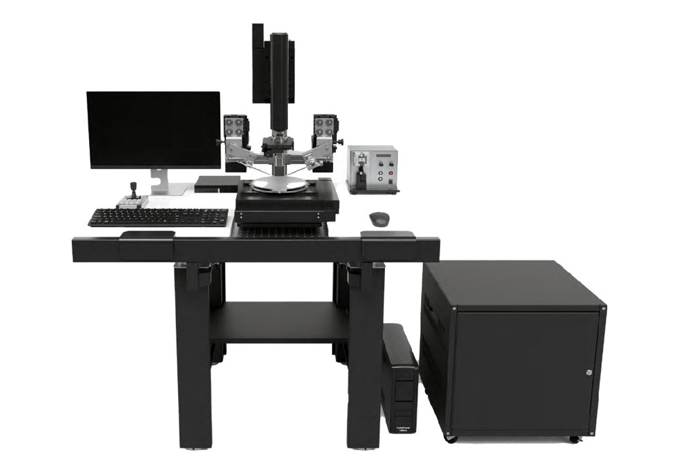
Fast, Precise Ex Situ Lift-Out with Tescan EXLO™ Tescan EXLO™ is designed for laboratories where TEM throughput, reproducibility, and cost efficiency matter. By moving specimen lift-out outside the FIB-SEM, EXLO™ keeps valuable beam time focused on milling, while parallelizing lamella transfer and grid mounting. The result: higher output, lower cost per specimen, and a more efficient workflow.
CATEGORY
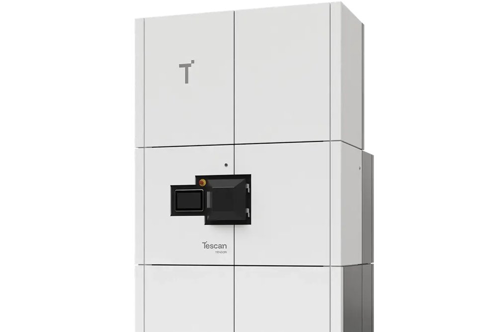
Nanoscale Insights with Tescan TENSOR™ Analytical STEM Tescan's TENSOR™ is the first 4D-STEM platform built for intuitive multimodal nanocharacterization, combining structural, morphological, and chemical insights in every scan. Real-time data processing, automation, and scripting support make it ideal for both routine and advanced research
CATEGORY