By General Nanotechnology, LLC
Accepts images from a broad range of AFM and similar scanning probe systems. Displays the images in 2D, but all 3D information is available and 3D cross sections can be displayed and measurements taken. A basic tool for visualization, annotation and documentation.
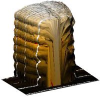
PROBE provides extensive tools for rendering, enhancing, analyzing and annotating 3D rendered images, some of which are described here: Dimensional Integrity: Dimensional axes may be shown or, as in this illustration, the axes may be embedded in the image. Dimensional integrity is maintained regardless of modifications to the image. Presentation Modes: 3D Height and (adjustable) Light. 3D rendered images may be shown in any of four presentation modes. Even in the 2D mode, Z-axis information is retained and shown. 2D Height Map; 3D Height; 3D Line Drawing. Image Rotation: Four image presentation modes are shown. A 3D image in any presentation mode can be rotated a full 360 degrees about both the X and the Y axes. Zoom: Selected areas of interest within images can be zoomed to enlarge features. The adjustable zoom window can be moved within the full image. Color Mapping: Images may be color mapped in several modes. Shown is Spectral mode, using multiple colors to indicate Z dimensions. The image can also be color mapped in Topographic mode, where color bands are delineated, as in a contour map. Gradation maps a single color, using brightness to indicate Z. Range of color (or brightness) can be selected and applied to a selected range of the Z data histogram. Relative Z dimensions may be color coded by color mapping. Filtering: Extensive filtering facilities are available to accentuate or enhance selected features or to reduce noise. Illustrated is a 2DFFT enhancement of a section of the upper edge of an etched hole. Other available filtering tools include: Smoothing, Sharpening, LaPlace, Median, Least Squares, Sobel, Mean Value, Three-Point Leveling, plus a 5 X 5 user-definable filter matrix. Measuring Tools: Cross sectional slice showing profile. Clicking within the profile window activates measuring tools. Extensive measurement capabilities are provided, using the ruler tools. Up to four Measurement Tables can be created from a single cross sectional window. Each table includes d(x), d(y), d(z), d(xy), d(xyz) and the angle between any two selected points. 3D Renderings from Optical Images: Stacks of 2D images can be rendered as 3D volumes (left) and 3D height maps (right). Cross sectional tool shows relative elevations. Interferometric Images: PROBE can import image files from a wide variety of imaging sources, plus Excel and MathCad files. Probe can accept interferomic and SPM image data from Veeco, and other interference and SPM microscope manufacturers, and from specialty imaging systems such as FRT. Annotation: The Select and Draw tools provide a full capability to add data in text and graphics to the images developed with Probe. Using the SuperClip module, composite images such as this can be captured and saved in all standard graphic file formats. Locating Tools: This group of tools provides extensive capabilities for identifying objects for further processing. Level Setting Tool: This tool allows setting profile level to accentuate features. Probe includes many additional enhancement, analysis and annotation tools.
CATEGORY
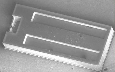
The basic nanotool™ V series consists of a Digital Instruments footprint MEMS cantilever platform capable of many new functions and features. These features include the ladder like structure down the middle of the part. The ladder is a thermal force generator and drives the two long arms which go up the sides of the part to clamp the tip platform and immobilize or provide higher spring constant to the mechanical action of the tip and primary cantilever. Additionally, the arms themselves may be covered by special silicon covers to form sealed continuous channels (see below bottom row right). These channels are fed from two large cavities under the main body by gas, vacuum or fluid. Furthermore a series of secondary cantilevers as can be seen just underneath the tip assembly (on the right upper row below) provide precise spring constant change as the primary cantilever is moved back and into them a micron or more. Many other new features are incorporated into the design and may be announced in the coming year as they reach production readiness. The back of the nanotool™ V series reveals the Dual Fluid or Gas chambers feeding the upper channel arms arranged around the tip platform. In this example the part has relatively weak secondary cantilever arms arranged just behind the tip platform to provide increased spring tension for conductive probing of subject materials. Nanotool™ V series are also available without any rear obstructions, beams or secondary cantilevers but in which the action of the clamping channel arms serves by design to make the appropriate spring constant change in the tip platform behavior. Inquire at GN to see if the special needs of your application can be supported by the nanotool™ Series of active cantilevers! Specific styles will be sampled at introductory prices to qualified companies and researchers.
CATEGORY
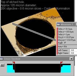
Optical micrscope imaging Damaged top of etched hole, 135 micron dia., showing etch damage Imaging and Analysis Software PROBE 3D renders 3D images from data files generated by Atomic Force Microscopes, Interferometric Microscopes, Scanning Probe Microscopes, Sequentially focused 2D optical images. PROBE can also import data files from Excel and MathCad and display the data as three-dimensional images. Regardless of the data source, the 3D image can be dynamically enhanced, analyzed and annotated using PROBE's extensive tools. It can then be filed, exported to documents or shared over networks with other PROBE users. PROBE runs on Windows NT (4.0), Windows 95/98, Win 2000, and XP operating systems. Its intuitively structured graphic user interface incorporates standard Windows features.
CATEGORY
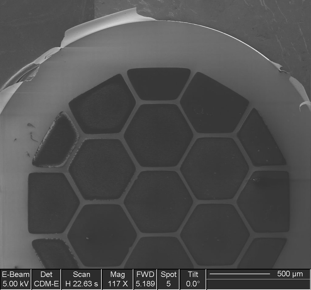
GN has created the first suspended membrane sample substrates on hexagonal silicon frames! Greater then 2 millimeters of nearly clear aperture let's you image and measure larger structures with all the advantages of suspended membrane technology! Nanotool™ TEM is a set of nine 50 nanometer thick Silicon Nitride low stress films on a silicon frame for mounting samples for Transmission Electron Microscopy and other applications. Diamond Like Carbon films as thin as 20 nm and 100 micron substrates are available on special order. GN's proprietary membrane sample carriers are the first to offer a full 2 mm nearly silicon free aperture with centered central hexagonal cells each with 576 microns of diagonal suspended membrane!
CATEGORY
