By Photothermal Spectroscopy Corp.
Available for mIRage and mIRage-LS featurefindIR microIR Chemical-ID is an integrated O-PTIR database that automatically analyzes all user selected spectra within a PTIR Studio file and correlates them against a reference set of O-PTIR spectra in an integrated database and provides a tabular output. Features Include: - Users can import and additional reference spectra from other sources - A hit quality index (HQI) is reported for every measured spectrum. - An overlay is displayed between the measured and reference spectra. - Color coding of common spectra is available for visual support Includes multiple common polymer datasets
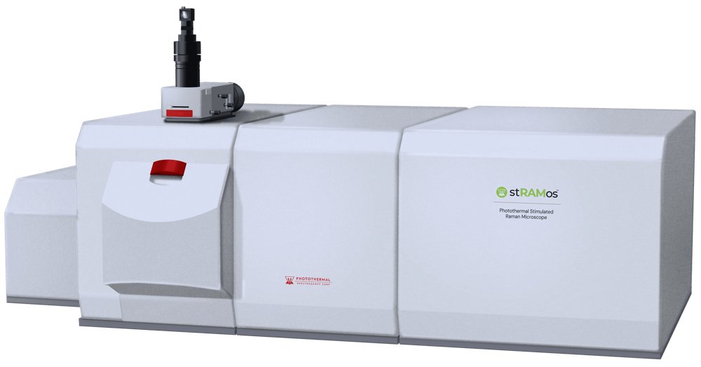
Sub-300nm Photothermal Stimulated Raman Microscope. Powered by PhotoThermalSRS™ Highest sensitivity measurements - 10x improvement over SRS ≤300nm Raman chemical spatial resolution High speed chemical imaging for live cells and molecular dynamics The stRAMos Photothermal Stimulated Raman Microscope uses the PhotoThermalSRS™ (PT-SRS) technique to deliver the combined highest measurement sensitivity and spatial resolution for chemical imaging in a single system with high imaging speeds and advanced microscopy characterization capabilities. The stRAMos microscope also uniquely combines many optional multimodal capabilities into a single system. stRAMos supports, the combination of PT-SRS with Optical Photothermal Infrared Spectroscopy (O-PTIR), widefield Epi Fluorescence imaging and spontaneous Raman spectroscopy, enabling the most thorough chemical characterization of your sample. stRAMos with its multimodal capabilities provides the most comprehensive chemical imaging and spectroscopy characterization for a wide range of life science applications including both dry and live cell imaging, biomolecular dynamics of proteins, lipids and other biomolecules. Hyperspectral Chemical Imaging in Live Cells PT-SRS enables full hyperspectral imaging across both the C–H and fingerprint regions, providing rich chemical contrast without labels. By capturing spectra at every pixel, researchers can distinguish subtle but critical molecular variations within live cells. This capability goes beyond single-wavelength imaging, allowing chemical fingerprints to be mapped across entire populations of cells. The result is a comprehensive view of cellular composition and dynamics — revealing heterogeneity, metabolic states, and molecular organization that would otherwise remain hidden. Hyperspectral imaging of live yeast cells from 2800cm -1 to 3017cm -1 High Speed, Label Free Imaging for Molecular Dynamics By enabling imaging at multiple frame rates than conventional techniques, stRAMos makes it possible to study biologically and chemically significant, molecular events. Many biomolecular dynamics—such as lipid transport, protein conformational changes, or membrane reorganization—occur on timescales where conventional imaging methods provide limited insight. With its unique ability to sensitively capture vibrational signals without labels, stRAMos bridges this gap, revealing dynamic processes that unfold over milliseconds to seconds. This opens new opportunities to monitor molecular reorganization, track metabolic activity, and investigate functional changes in living systems with unmatched clarity. High speed single wavelength imaging of cells. Imaging of lipids measured at 2850cm -1. Volumetric chemical sectioning with submicron resolution stRAMos enables 3D chemical mapping of cells with unprecedented clarity. Unlike conventional volumetric methods, which often compromise resolution, stRAMos delivers true submicron sectional imaging. This allows researchers to resolve fine structural and biochemical details within cells, revealing organization and molecular composition layer by layer. By combining chemical specificity with high-resolution sectioning, stRAMos provides a powerful new way to study cellular architecture and molecular distribution in three dimensions — without labels or staining. 3D Volumetric imaging of lipids and proteins in a yeast cell. Video of biomolecules measured at multiple depths at the fixed wavenumber of 2850cm-1. Image depth varies down to 17µm. Image slightly speeded up. Multimodal imaging with correlative O-PTIR and Fluorescence Microscopy The stRAMos system uniquely combines optional multimodal capabilities into one platform. In a combined system, the stRAMos supports PhotoThermalSRS with Optical Photothermal Infrared Spectroscopy (O-PTIR), Widefield Epi Fluorescence microscopy and spontaneous Raman spectroscopy.
CATEGORY
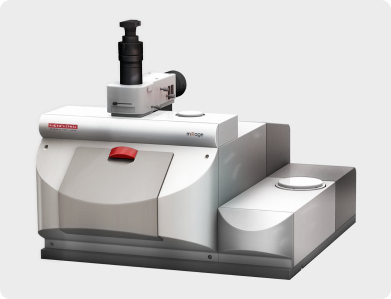
The mIRage platform is modular microscope that delivers high performance submicron IR spectroscopy and imaging with a range of options to address broad application flexibility. Non-contact reflection IR measurement with FTIR-quality spectra and no artifacts Fast, easy to use with little to no sample preparation and spectroscopy in seconds Overcomes the limitations of Raman spectroscopy About mIRage mIRage overcomes the IR diffraction limit using Optical Photothermal Infrared (O-PTIR) Spectroscopy, in which a tunable pulsed mid-IR laser induces a photothermal response from the sample surface. This thermal response is measured using a colinear visible probe laser focused on the sample. Spatial resolution is determined by the diffraction limited spot size of the visible laser (around 500 nm), therefore independent of the IR wavenumber, unlike traditional diffraction limited techniques. Transmission FTIR quality spectra in non-contact reflection mode As mIRage is an optical microscope based platform, measurements are collected quickly and easily, without the need for sample contact, unlike ATR spectroscopy. Additionally, O-PTIR provides spectra comparable to FTIR, ideal for database searches, without the dispersive artifacts observed in ATR. By operating in reflection mode, the need for thin samples is also eliminated. Simultaneous IR and Raman spectral searching, complementary and confirmatory Traditionally IR and Raman spectral searching has been done separately and the data has been analyzed separately. Now, with Wiley’s recent release of the KnowItAll™ multi-technique of simultaneous searching, the hit list information is displayed in a graphical 2D scatter plot. This multi-dimensional approach plots the HQI of the unknown IR spectrum against the HQI of the unknown Raman spectrum dramatically speeding up data analysis. This greatly simplified display of spectral matches in graphical rather than tabular form greatly improves the accuracy of the identification. IR search results can confirm Raman search results and vice-versa, all simultaneously. IR+Raman: Same time. Same spot. Same resolution. The world’s first simultaneous IR+Raman microscopy system is a unique dual modality platform with all the advantages of an O-PTIR and Raman microscope combined. O-PTIR, optical photothermal microscopy, using a visible probe laser, is combined with Raman for applications in polymers, defect analysis, microplastic detection and red blood cells. Spectra, line scans and 2D maps can now be collected from the same spot at the same time, opening up new research opportunities and a more thorough characterization of your sample. Wide range of applications mIRage provides unique data for numerous applications and material types, including: - Microplastics - Failure analysis - Microelectronics - Polymers and composites - Defect identification - Geological sciences - Forensics Microplastics The mIRage uniquely provides accurate measurement of a wide range of critical <20 µm microplastics that are poorly resolved by other IR and Raman techniques. Biopharma Integrating sub-micron chemical and morphological analysis into routine particle control strategies is now a reality with the mIRage platform, submicron multimodal IR microscope. MiRage provides routine analysis of submicron chemical and morphological analysis of small particulate matter, overcoming the limitations of other spectroscopy techniques. Semiconductor mIRage provides the ability to identify unknowns of small size using sub-micron O-PTIR or simultaneous IR+Raman.
CATEGORY
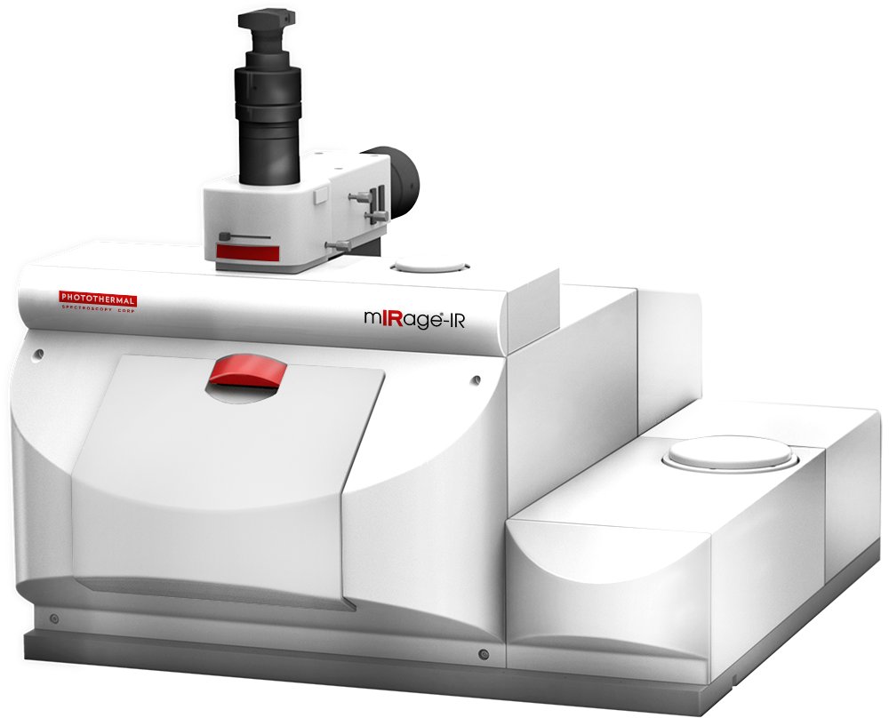
Submicron IR spectroscopy with FTIR quality data. Why settle for FTIR? Step up to submicron O-PTIR. Submicron resolution spectroscopy and Imaging. Non-contact reflection IR measurement with FTIR-quality spectra and no artifacts. Fast, easy to use with little to no sample preparation and spectroscopy in seconds. featurefindIR for automated workflows with integrated Micro Chemical ID. The mIRage-IR delivers cutting-edge submicron infrared (IR) spectroscopy and chemical imaging using proprietary Optical Photothermal IR (O-PTIR) technology — at a price point competitive with traditional FTIR/QCL microscopes. Designed for researchers and scientists seeking better IR spatial resolution while maintaining FTIR transmission/ATR-likespectra, and improved ease of use through easy reflection mode operation, mIRage-IR opens the door to next-generation performance in the same budget class. The mIRage-IR is ideal for the study of particulates, like microplastics, (bio)pharmaceutical powders and sub-visibles, materials science/polymers, through to environmental aerosols and industrial failure analysis/contaminants. As mIRage-IR is an optical microscope-based platform, measurements are collected quickly and easily, in reflection mode making it a very easy “point and shoot” process, without the need for sample contact, unlike FTIR-ATR spectroscopy and other contact- based IR techniques. FTIR Transmission/ATR-like spectra in non-contact reflection mode: O-PTIR provides submicron IR resolution spectra comparable to FTIR, thus existing custom or commercial FTIR databases are fully compatible. While operating primarily in reflection mode, O-PTIR delivers spectra that are free of dispersive scattering artefacts that have traditionally limited direct IR (FTIR/QCL) techniques in reflection mode. Easier sample preparation and greater sample flexibility: By operating in reflection mode, minimal to no sample preparation is required, because sample thickness or surface roughness issues are eliminated. Standard glass slides and non-coated filters (like polycarbonate, nitrocellulose etc) are fully compatible on the mIRage-IR, providing broader application flexibility. FeaturefindIR: Detect, Select, Measure, Identify. featurefindIR can accurately and automatically measure and chemically identify particles and micro-plastics from submicron to mm’s in size, overcoming the limitations of traditional direct IR (FTIR/QCL) and Raman microscopy techniques. Wide range of applications: mIRage-IR provides unique data for numerous application and material types, including: - Microplastics - Failure analysis - Microelectronics - Polymers and composites - Defect identification - Geological sciences - Forensics. 5 Reasons to choose mIRage-IR as your next IR microscope: Achieve Sub Micron Resolution on your samples - mIRage-IR achieves sub-micron spatial resolution – more than 10× higher than any FTIR microscope. - By detecting the photothermal IR absorption from a 0.5 microns visible probe spot size, the spatial resolution is always independent of IR wavelength. Analyze without sample preparation - O-PTIR is always non-contact and works directly on flat, rough, transparent and opaque samples without compression, cutting, or complex preparation. - Obtain rapid, reproducible measurements on fragile, valuable, or hydrated specimens. - Even use glass substrates since mIRage-IR operates in reflection mode using a visible light probe to detect IR absorption indirectly. Capture artefact free FTIR transmission/ATR-like spectra in reflection mode - By detecting O-PTIR absorption indirectly with the visible probe, mIRage-IR eliminates scattering artefacts and morphology dependent distortions. - Clean, flat baseline spectra ensure accurate materials identification from FTIR libraries. - mIRage-IR’s sensitive IR spectra enable repeatable assignment and component grouping from each spectrum. Accelerate workflows with combined O-PTIR and fluorescence - Correlate label-free chemical information from O-PTIR with fluorescence and autofluorescence based identification of specific molecules or structures with sub-micron resolution. - Integrate chemical and biological insights seamlessly, enhancing standard life science workflows and track labelled and autofluorescent particles including micro and nano plastics. Obtain highest sensitivity and highest resolution IR chemical imaging! - Capture the most detailed, label-free molecular information with enhanced sensitivity and spatial clarity, enabling analysis of heterogeneous samples with small or complex features. - Sub-micron resolution minimises signal averaging and the absence of scattering artifacts ensures reproducibility. - Acquire multi-wavelength image sets in under 5 minutes with each image individually mapping the distribution of specific species on your sample. Upgradeable to co-located fluorescence microscopy for higher productivity measurements. Do you have autofluorescent defects that you need to detect? Our co-located fluorescence and O-PTIR imaging camera easily detects these types of defects enabling higher measurement productivity. Co-located fluorescence and submicron IR enables analysis of micro-electronics devices and many other applications areas. Microplastics applications: Detect, Select, Measure, Identify. The mIRage-IR uniquely provides accurate measurement of a wide range of critical <20 µm microplastics that are poorly resolved by other IR and Raman techniques. Pharma and Biopharma applications: Detect, Select, Measure, Identify. Integrating submicron chemical and morphological analysis into routine particle control strategies is now a reality with the mIRage platform, submicron multimodal IR microscope. miRage-IR provides routine analysis of submicron chemical and morphological analysis of small particulate matter, overcoming the limitations of other spectroscopy techniques. Failure analysis applications: Detect, Select, Measure, Identify. mIRage-IR provides the ability to identify unknowns of small size using submicron O-PTIR or simultaneous IR+Raman.
CATEGORY
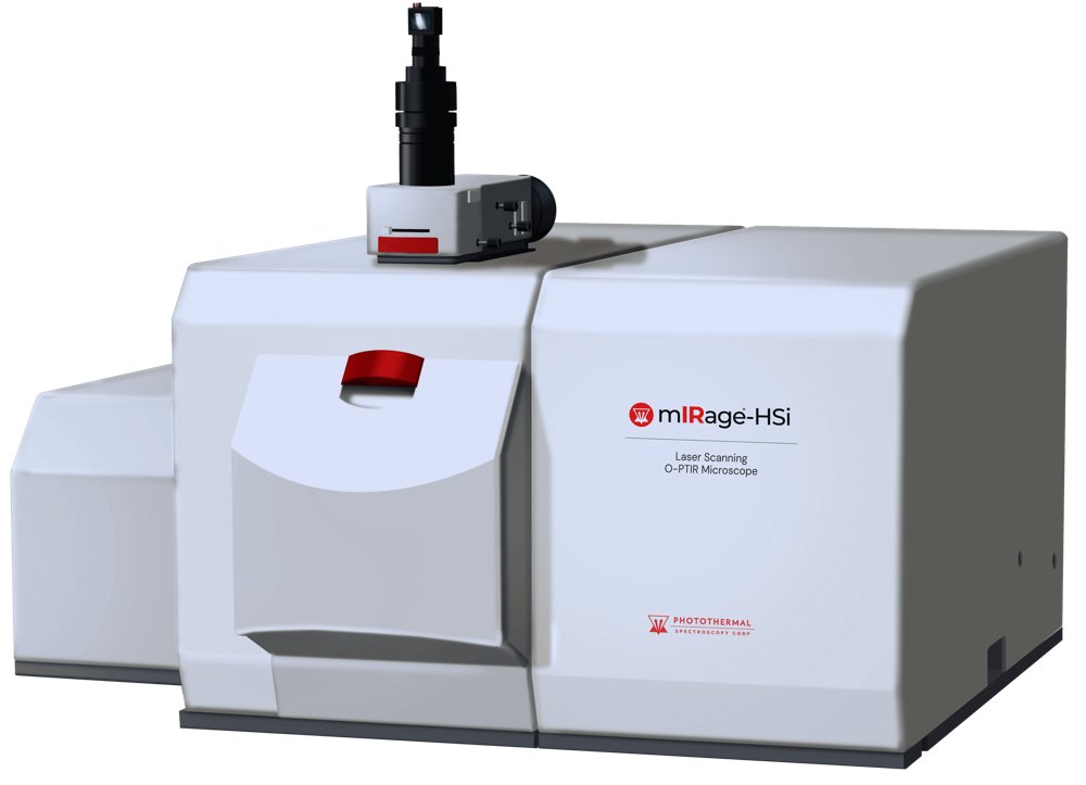
Sub-500nm laser scanning O-PTIR microscope for ultra-high-speed imaging. With co-located fluorescence imaging and Raman spectroscopy. 30x faster O-PTIR: Hyperspectral imaging in minutes, single wavelength imaging in seconds Sub-500nm IR chemical spatial resolution Multi-modal correlative fluorescence imaging and Raman spectroscopy Next-level performance: Laser scanning O-PTIR The new mIRage-HSi is the next evolution of Optical Photothermal Infrared (O-PTIR) spectroscopy and imaging, delivering the highest performance sub-500nm IR imaging and FTIR transmission/ATR-like spectroscopy but now with breakthrough speed reducing hyperspectral imaging time from hours to minutes and single frequency images from minutes to seconds. This dramatic increase in imaging speed extends O-PTIR technology to dynamic studies, higher speed contaminant detection, and automated submicron particle identification. Whether for microplastics, pharma, life science, failure analysis, or materials science, mIRage-HSi delivers faster, more automated, and more productive O-PTIR workflows. Improved O-PTIR imaging measurement speed mIRage-HSi: 16 seconds/image 30X speed improvement Tissue section: Imaged 1655 cm-1 (protein) FOV: 75x75 µm, 200 nm pixel size Automated multi-wavelength imaging Image sequence & RGB overlay PTIR RGB overlay provides an automated measurement method for user selectable wavenumbers with resulting RGB overlay The RGB overlay example shows 3 distinct wavenumbers representing: 1080cm -1 – Nucleic acid 1655 cm -1 – Proteins 1738 cm -1 – Lipids Each wavenumber took 16 seconds. Preview mode: real-time chemical imaging for ROI significant EoU and productivity improvement Preview mode is a lower resolution, high speed chemical imaging preview of a selected area. It provides a significant enhancement in measurement productivity for locating the feature of interest. Move, Locate, Image Quickly identify the relevant ROI for defects, contaminants, microplastics over an area greater than the objective FOV.
CATEGORY
