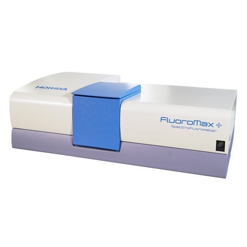
Steady State and Lifetime Benchtop Spectrofluorometer The FluoroMax®Plus represents HORIBA's industry-leading fluorometer performance in a convenient, affordable, easy-to-use benchtop model. The FluoroMax family, with its unique all reflective optics and photon counting was the first to bring the sensitivity of a modular fluorometer to a tabletop fluorescence instrument, in the 1990's. It’s a compact spectrofluorometer
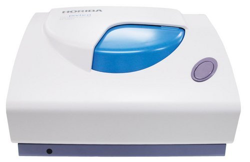
Nanoparticle Analyzer. A highly advanced analyzer solves the mysteries of the nano-world. A single device analyzes the three parameters that characterize nanoparticles: particle size, zeta potential, and molecular weight. Nanotechnology research and development is a continuously evolving effort to control substances at the atomic and molecular level in order to achieve new and better materials and products. The miniaturization of components – that is, control at the nano-level – is necessary to achieve faster, higher-performance devices and functions and to reduce energy consumption. Nanotechnology has come to play a key role in wide-ranging fields that affect our daily lives, including food, cosmetics, and the life sciences. Clear and simple multi-parameter analysis of nanoparticles! Three analyzers in a single compact body deliver high-sensitivity, high-accuracy analysis of each measurement parameter. Manufacturing Company: HORIBA, Ltd. Particle Size Measurement Range 0.3 nm to 10 µm: The SZ-100V2 Series measures particle size and particle distribution width by dynamic light scattering (DLS). Analysis across a wide range of sample concentrations: Measurement of samples ranging from low ppm-order concentrations to high-concentration samples in double-digit percentages is possible. Accepts commercially available sampling cells. Analysis of small-volume samples is also possible. Zeta Potential Measurement – 500 to + 500 mV: Analysis of sample volumes as small as 100 μL using HORIBA-developed microelectrophoresis cells. Use the value of zeta potential to predict and control dispersion stability. High zeta potential magnitudes indicate a stable dispersion, useful for formulation work. Molecular weight 1 × 10 3 to 2 × 10 7 Da: Absolute molecular weight (Mw) and the second virial coefficient (A 2) are obtained by performing static light scattering measurement as a function of sample concentration and preparing Debye plots. The SZ-100V2 Series applies sophisticated intelligence and learning capability to rapidly determine nanoparticle properties! - Since the SZ-100V2 Series analyzer covers a wide sample concentration measurement range, sample dilution and other preprocessing is nearly eliminated. - The use of a dual optical system enables measurement of high-concentration samples such as slurry and ink pigments as well as low-concentration proteins and polymers. - A single device analyzes the three parameters that characterize nanoparticles: particle size, zeta potential, and molecular weight. - HORIBA-developed cells for zeta potential measurement prevent sample contamination. Simple analysis by means of ultra micro-volume dedicated cells (volume as low as 100 μL). Suitable for analysis of dilute samples. - HORIBA-developed electrode for zeta potential cell made from carbon material, the material is not corroded by high salt samples such as saline. SZ-100-S2 Measurement Specifications: Model: SZ-100-S2 and SZ-100-HS2 (particle size and molecular weight measurement only) Measurement principles: Particle size measurement: Dynamic Light Scattering; Molecular weight measurement: Debye plot method (static scattered light intensity) Measurement range: Particle size: 0.3 nm to 10 μm; Molecular weight: 1000 to 2 x 10 7 Da (Debye plot); 540 to 2 x 10 7 Da (MHS Equation) Maximum sample concentration: 40 wt% Particle size measurement accuracy: Measurement accuracy of ±2% for NIST traceable polystyrene latex 100 nm spheres (not including variation in the standard particles themselves) Measurement angles: 90° and 173° (automatic or manual selection) Cells: Cuvettes Measurement time: Approx. 2 min. under ordinary conditions (from the start of measurement to the display of results for particle size measurement) Required sample volume: Minimum volume of 12 μL to 1000 μL (differs depending on cell material) Usable liquids: Water, ethanol, organic solvents SZ-100-Z2 Measurement Specifications (Particle size and molecular weight measurement specifications are the same as for the SZ-100-S2.): Model: SZ-100-Z2 and SZ-100-HZ2 (with zeta potential measurement unit) Measurement principle: Zeta potential measurement: Laser Doppler electrophoresis Measurement range: -500 to +500 mV Size range suitable for measurement: Minimum 2.0 nm, Maximum 100 μm Measurement conductivity range: 0 to 20 S/m; Recommended sample conductivity range: 0 to 2 S/m Maximum sample concentration: 40 wt% Cells: Dedicated cell with electrodes Measurement time: Approx. 2 min. under ordinary conditions Required sample volume: 100 μL Carrier fluids: Water Analyzer Specifications (SZ-100-S2 and SZ-100-Z2): Measuring unit optical system: Light source: Diode pumped frequency doubled laser (532 nm, S2 / Z2 10 mW, HS2 / HZ2 100 mW); Detectors: Photomultiplier tubes (PMT) Laser classification: Class I Operating temperature and humidity: 15 - 35 °C, RH 85% or less (no condensing) Holder temperature control temperature settings: 0 - 90 °C (up to 70 °C for cells with electrodes and plastic cells) Purging: Dry gas purge port tube connection is possible. Power supply: AC 100 - 240 V, 50/60 Hz, 150 VA Dimensions: 528 (W) x 385 (D) x 273 (H) mm (excluding protrusions) Weight: 25 kg Personal computer: Windows computer with one available USB port Interface: USB 2.0 (between measuring unit and PC)
CATEGORY
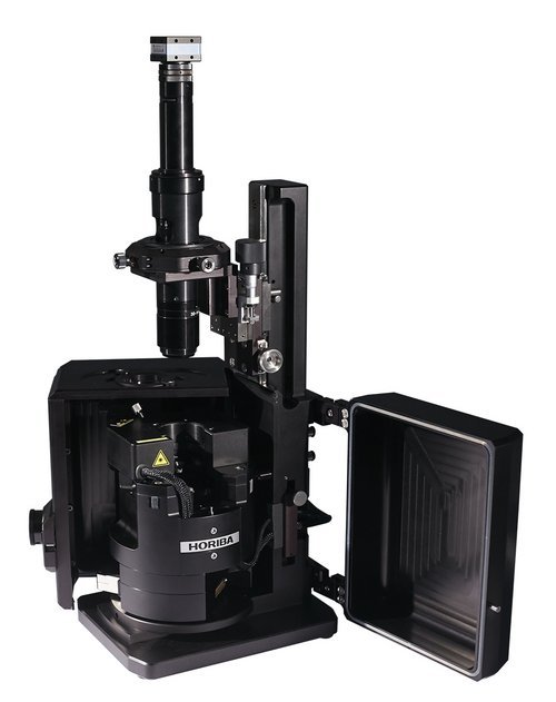
Automation of operation / Ease of use Fully automated laser to photodiode alignment (no user click-on-spot is required!). Exchanging probes has never been so easy before! Fully automated configuration for the most common AFM modes. Extremely fast system adjustment before starting measurements! Resolution / Stability / Accuracy One single 100µm scanner gets large scans as well as goes down to molecular or atomic resolution. No vibration isolation is required for standard measurements. The lowest noise closed loop sensors make it possible to have them on even during molecular resolution imaging and get the most accurate results. Fast scanning Scanner resonant frequencies 7kHz in XY and 15kHz in Z are the highest in the AFM industry today. Optimized scanner control algorithms makes it possible to scan much faster than ever before! All SPM modes included plus Nanolithography with no additional units and costs Kelvin Probe Microscopy, Piezoresponse Force Microscopy, Nanolithography and Nanomanipulation are all included in the basic package! Flexibility to upgrade to AFM-Raman The SmartSPM was designed from the ground up for easy and appropriate coupling with Raman systems. Measuring Modes - Contact AFM in air; - Contact AFM in liquid (optional); - Semicontact AFM in air; - Semicontact AFM in liquid (optional); - True Non-contact AFM; - Dynamic Force Microscopy (DFM, FM-AFM); - Dissipation Force Microscopy; - Top Mode; - Phase Imaging; - Lateral Force Microscopy (LFM); - Force Modulation; - Conductive AFM (optional); - I-Top mode (optional); - Magnetic Force Microscopy (MFM); - Kelvin Probe (Surface Potential Microscopy); - Single-pass Kelvin Probe; - Capacitance Microscopy (SCM) - Electric Force Microscopy (EFM); - Single-pass MFM/EFM (“Plane scan”); - Force curve measurements; - Piezo Response Force Microscopy (PFM); - PFM-Top mode; - Nanolithography; - Nanomanipulation; - STM (optional); - Photocurrent Mapping (optional); - Volt-ampere characteristic measurements (optional); - Shear-force Microscopy with tuning fork (ShFM); - Normal Force Microscopy with tuning fork. SmartSPM Scanner and Base Sample scanning range:100 µm x 100 µm x 15 µm (±10 %) Scanning type by sample:XY non-linearity 0.05 %; Z non-linearity 0.05 % Noise:< 0.1 nm RMS in XY dimension in 200 Hz bandwidth with capacitance sensors on;< 0.02 nm RMS in XY dimension in 100 Hz bandwidth with capacitance sensors off;< 0.04 nm RMS Z capacitance sensor in 1000 Hz bandwidth Resonance frequency:XY: 7 kHz (unloaded);Z: 15 kHz (unloaded) X, Y, Z movement:Digital closed loop control for X, Y, Z axes; Motorized Z approach range 18 mm Sample size:Maximum 40 x 50 mm, 15 mm thickness Sample positioning:Motorized sample positioning range 5 x 5 mm Positioning resolution:1 µm AFM Head HE001 Laser wavelength: 1300nm; No registration laser influence on biological sample; No registration laser influence on photovoltaic measurements. Registration system noise: <0.03nm. Fully motorized: 4 stepper motors for cantilever and photodiode automated alignment. Free access to the probe for additional external manipulators and probes; Top and side simultaneous optical access: planapochromat objectives 10x, NA=0.28 and 20x, NA=0.42 respectively. AFM Head HE002 Laser wavelength: 1300nm; No registration laser influence on biological sample; No registration laser influence on photovoltaic measurements; Registration system noise: <0.1nm; Fully motorized: 4 stepper motors for cantilever and photodiode automated alignment; Free access to the probe for additional external manipulators and probes; Top and side simultaneous optical access: with planapochromat objectives, Side objective up to 100x, NA=0.7 Top objective 10x, NA=0.28 simultaneously; With HE002 Contact and Semicontact AFM in liquid are impossible. Optical microscope Numerical aperture: up to 0.1; Magnification: on 19" monitor with 1/3" CCD from 85x to 1050x; Horizontal field of view: from 4.5 to 0.37 mm; Manual detent zoom: 12.5x (motorized zoom optional); Stand and coarse/fine focusing unit; Capability to use planapochromat objectives 10x, NA=0.28 and 20x, NA=0.42 and 100x, NA=0.7 (depends on AFM head); Liquid cell (optional) Sample size: 2mm thickness, 25mm diameter; Sample positioning range: 5x5mm; Positioning resolution: 1um; Cell size: 40x40x12mm; Volume of liquid: 3ml; Capability of liquid exchange; Autoclave and ultrasonic cleaning of cell parts. Liquid cell with temperature control (optional) Sample size: 2mm thickness, 25mm diameter; Heating: up to 60°C; Cooling: below room temperature down to 5°C; Sample positioning range: 5x5mm; Positioning resolution: better than 1.5um; Cell size: 40x40x12mm; Volume of liquid: 3ml; Capability of liquid exchange; Autoclave and ultrasonic cleaning of cell parts. Conductive AFM unit (optional) Current range: 100fA ÷ 10uA; 3 current ranges (1nA, 100na and 10uA) switchable from program; Voltage range: -10 ÷ +7V; RMS current noise: less than 60fA for 1nA range. Conductive to Kelvin mode switchable from program. Combined Shear-force and Normal force tuning fork holder (optional) Compatible with tuning forks with free resonant frequency 32,768 kHz; Two types of PCBs for tuning fork fixation for shear-force and normal force operation; Piezoceramic excitation of tuning fork vibration; Integrated preamplifier for low noise; Compatible with 100x side objective for TERS\TEPL measurements; Compatible with 10x top objective. Nanoindenter unit (optional) Maximum load: 5mN. Load noise floor: less than 100nN. Displacement noise floor: less than 0.2 nm. Compatibility with optical systems No interference with optical imaging due to infrared laser; Upgradeability to OmegaScope for spectroscopic, photovoltaic, SNOM and TERS & TEPL operation. Protection enclosure with stand for optical microscope (optional) Acoustic isolation: 30dB; Electrostatic shielding; Inlet and outlet fittings for environmental control connection; Focusing with micrometer screw ± 6.5mm; Micrometer reading: 10 um; Focusing sensitivity: better 1um; Fast service lift: range up to 98mm; XY positioning: ± 2mm; Optional fine focusing for 100x objective: Fine focusing 0.2mm; Micrometer reading 0.5 um; Focusing sensitivity better 0.1um; Humidity control system (optional) Relative humidity range: 10-85%; Relative humidity stability: ±1%; Software Automatic alignment of registration system; Automatic configuration and presetting for standard measuring techniques; Automatic cantilever resonance frequency adjustment; Capability to work with force curves; Macro language Lua for programming user functions, scripts and widgets; Capability to program controller with DSP macro language in real time without reloading control software; Capability to process images in coordinate space including making cross-sections, fitting and subtracting of polynomial surface up to 12 degree; FFT processing with capability to treat images in frequency space including filtration and analysis; Nanolithography and nanomanipulation; Processing up to 5000x5000 pixel images.
CATEGORY
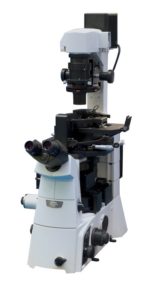
Extraordinary productivity and easy operation CombiScope is equipped with the fully motorized cantilever holder and photodiode positioning that provides the automated click-on-a-button laser-to-tip alignment. This option dramatically simplifies the entire system adjustment process and provides the highest level of system adjustment reproducibility. In addition, after you installed a new cantilever of the same or even different type, the same spot (within a few microns repeatability) on your sample surface can be easily found and scanned without any extra searching steps. Top level scanner The CombiScope utilizes the closed loop, high-dynamics, 3-axis piezo-nanopositioning scanner from the leader in precision motion control, Physik Instrumente (PI). The top-level scanner is the heart of the system which enables it to achieve very high levels of linearity, highest possible stiffness and extremely high precision motion. 1300 nm AFM laser The use of 1300nm AFM laser eliminates any interference with VIS light-sensitive biological and semiconductor samples. It also makes it possible to perform simultaneous AFM and fluorescence or Raman scattering measurements without any crosstalk for most popular UV-VIS-NIR (364-830 nm) excitation lasers. Integration with optics Besides integrated inverted optical microscope such as Nikon Eclipse Ti-U and Olympus IX-71 with Phase Contrast and DIC, the CombiScope can be equipped with the head which provides the top and side simultaneous optical access with planapochromat objectives (100x, NA=0.7 and 10x, NA=0.28 respectively). This option opens up the way to combine the upright and transmission configurations to study transparent as well as non-transparent samples with optical, Raman and scanning probe microscopy techniques. Solutions for working in liquid The standard CombiScope's sample holders accommodates all common sample substrates, including slides, cover slips and 35mm Petri dishes. The specially design liquid cell with heating and liquid perfusion capabilities enables for biological samples to be delicately maintained in their physiological environment and at temperatures up to 60°C. All operating modes in one single instrument The CombiScope comes with all modern AFM operating modes in one single instrument, without any extra costs and units, including such application-specific modes as force and electric nanolithographies, piezoelectric force microscopy (PFM), Kelvin Probe Microscopy and frequency modulation AFM (dynamic force microscopy with built-in PLL). In addition, the scanning tunneling microscopy (STM) head and Conductive AFM unit operating in the range 100fA ÷ 10uA (with 1nA, 100na and 10uA subranges software switchable and current noise of 60fA RMS for 1nA subrange) and near-field optical microscopy (SNOM) head are available as the options. Such exceptional versatility of the instrument makes it a perfect solution for nanoscience. Measuring Modes Contact AFM in air; Contact AFM in liquid (optional); Semicontact AFM in air; Semicontact AFM in liquid (optional); True Non-contact AFM; Dynamic Force Microscopy (DFM, FM-AFM); Dissipation Force Microscopy; Top Mode; Phase Imaging; Lateral Force Microscopy (LFM); Force Modulation; Conductive AFM (optional); I-Top mode (optional); Magnetic Force Microscopy (MFM); Kelvin Probe (Surface Potential Microscopy); Single-pass Kelvin Probe; Capacitance Microscopy (SCM) Electric Force Microscopy (EFM); Single-pass MFM/EFM (“Plane scan”); Force curve measurements; Piezo Response Force Microscopy (PFM); PFM-Top mode; Nanolithography; Nanomanipulation; STM (optional); Photocurrent Mapping (optional); Volt-ampere characteristic measurements (optional). Measuring Modes in liquid with AFM Head HE001 and Liquid cell Contact AFM; Semicontact AFM; TopMode; Phase Imaging; Lateral Force Microscopy (LFM); Force Modulation; Force curve measurements; Nanolithography; Nanomanipulation. Scanner and Base Sample scanning range: 100 µm x 100 µm x 20 µm (±10 %) Scanning type by sample: XY non-linearity 0.05 %; Z non-linearity 0.05 % Noise: 0.1 nm RMS in XY dimension in 100 Hz bandwidth with capacitance sensors on; 0.02 nm RMS in XY dimension in 100 Hz bandwidth with capacitance sensors off; < 0.1 nm RMS Z capacitance sensor in 1000 Hz bandwidth Resonance frequency: XY: 7 kHz (unloaded); Z: 15 kHz (unloaded) Base Manual sample positioning: range 25x25mm, positioning resolution 1um; Motorized SPM measuring head positioning: 1.6x1.6mm, positioning resolution 1um; Motorized approach: 1.3 mm; Sample holder for standard slides and cover glasses; Optional Sample holder: Maximum sample size: 50.8x50.8 mm, 5 mm height with capability to choose measuring area 25x25mm in any quadrant of 50.8x50.8 mm area or in center of sample. AFM Head HE001 Laser wavelength: 1300nm; No registration laser influence on biological sample; No registration laser influence on photovoltaic measurements. Registration system noise: <0.03nm. Fully motorized: 4 stepper motors for cantilever and photodiode automated alignment. Free access to the probe for additional external manipulators and probes; Top and side simultaneous optical access: planapochromat objectives 10x, NA=0.28 and 20x, NA=0.42 respectively. Liquid cell (optional) Manual sample positioning: range 15x15mm, positioning resolution 1um; Holder for Petri dish dia.35mm; Volume of liquid: 1.5-2.5 ml; Flow-through for liquid exchange: two tubes. Conductive AFM unit (optional) Current range: 100fA ÷ 10uA; 3 current ranges (1nA, 100na and 10uA) switchable from program; Voltage range: -10 ÷ +7V; RMS current noise: less than 60fA for 1nA range. Compatibility with inverted optical microscopes No interference with optical imaging due to infrared laser; Capability to install on: Nikon Ti-E, Ti-U, Ti-S, TE2000; Olympus IX-71, IX-81; Phase contrast, DIC and fluorescent techniques with native optical condenser; Upgradeability to TRIOS platform for spectroscopic and TERS operation. Optical microscope for standalone operation (optional) Numerical aperture: up to 0.1; Magnification on 19" monitor with 1/3" CCD: from 85x to 1050x. Horizontal field of view: from 4.5 to 0.37 mm Manual detent zoom: 12.5x Stand and coarse/fine focusing unit.; Capability to use planapochromat objectives: 10x, NA=0.28 and 20x, NA=0.42 and 100x, NA=0.7 (depends on AFM head); Software Automatic alignment of registration system; Automatic configuration and presetting for standard measuring techniques; Automatic cantilever resonance frequency adjustment; Capability to work with force curves; Macro language Lua for programming user functions, scripts and widgets; Capability to program controller with DSP macro language in real time without reloading control software; Capability to process images in coordinate space including making cross-sections, fitting and polynomial smoothing up to 8 degree; FFT processing with capability to treat images in frequency space including filtration and analysis; Nanolithography and nanomanipulation; Processing up to 5000x5000 pixel images.
CATEGORY
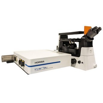
Fluorescence Lifetime Imaging Platform The InverTau™ platform is a laser scanning confocal unit for Fluorescence Lifetime Imaging (FLIM).InverTau is fully software controlled and designed to fit on the side port of an inverted fluorescence microscope. Build your own microscope system around the InverTau platform, or buy a complete system from HORIBA. Versatile solutions are always available with local support. Features: - Affordable laser scanning unit for FLIM studies - Fully computer-controlled confocal optics with the intuitive EzTime Image Software minimizes set up time and increases productivity - Simply switch from confocal laser scanning to widefield imaging with the addition of FLIMera - Real time video display up to 6 fps with InverTau and up to 30 fps with addition of FLIMera - Add InverTau to the side port of an inverted microscope or buy a complete solution from HORIBA - Intuitive software and automated optics minimize setup time and increase productivity InverTau Specifications: Modes: Single point, raster, bi-directional (xy) Scan speed: Up to 6 fps (bi-directional) at 512x512 Image resolution: 32x32 to 4k x 4k Digital zoom: 1FoV to 0.1FoV Attachment: To side port of inverted microscope (C-mount) Control software: EzTime™ Image software Optical ports: 2 on excitation, 2 on emission Attenuation filters: Steps from 0-6 OD ND Dichroic filters: 400 nm, 450 nm, 500 nm, 550 nm, 600 nm, 730 SP, 50/50 BS Emission filters: 400BP, 450BP, 500BP, 550BP, 600BP,440LP, 475LP, 530LP, 570LP, 630LP Pinhole: 10, 25, 50, 100, 200 μm (+ closed and fully open) Photon streaming: Stream to HDF5 Phasor analysis: Switchable with histogram Data analysis: Global up to 5 exponentials Key FiPho Electronics Specifications for FLIM: Lifetimes from <50 ps Deadtime: 5 ns TCSPC bin width: <15 ps Histogram Bin Depth: 8, 16 and 32-bit Independent Stops: 1 to 4 Maximum Start Rate: 100 MHz Maximum Rate/Stop Channel: 10 Mcps Operating Mode: Automatic Forward Timing Streaming Mode: Photon Streaming (Time-Tag) Acquisition and Analysis Macro Scripting: Yes PC Interface: USB 3.0 Software: EzTime™ Image
CATEGORY
