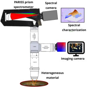
The PARISS Imaging Spectrometer is prism-based for the highest possible spectral sensitivity. Zoom magnification: From less than 1x to greater than 40x. Eliminates the need for multiple objectives Samples and specimens: Designed to work with heterogeneous samples in air, a petri dish, microscope slide, in water or in-situ Optics and objectives: Accommodates high numerical aperture (NA) long working distance objectives, commercial microscope objectives, camera, and long focal length telescope lenses for remote sensing.Spectroscopy modes: %reflection, %transmission, absorption fluorescence, luminescence… Wavelength range: prism-based imaging spectrograph. Operates from 360 – 920-nm depending on the spectrum camera and application optics.Mounts: Can be mounted on a column, microscope C-mount video port, a cart, tripod, or mast Illumination: LED brightfield, darkfield, refection, transmission, monochromatic fluorescence excitation Observed field-of-view: Coaxial side camera records the FOV live. Great for object targeting and documentation PARISS Specifications Weight: 1,250 g (Excluding a camera)Moving parts: None. Optimizes stability and reproducibility. Dimensions: 210 x 55 x 85 mm Wavelength dispersive element: The wavelength dispersive element is a prism with optical “power.” Concave and convex surfaces on the front and rear surfaces correct astigmatism, coma, and spherical aberration. (See Figure 2)Spectral range: 365 to ~920 nm or 400 to ~920 nm, depending on choice of camera. All spectra acquired simultaneously without order sorting filters Light throughput efficiency: Internal transmission ~90% from 450 to ~920 nm.Entrance slit dimensions: Standard 5 mm by 25-micron, widths of 50 and 100-micron are available in pre-aligned mounting assemblies.Spatial resolution at the sample: Depends on slit width and camera pixel size ~ 0.6 micron by ~0.6 micron with 40x magnification typical. Nanoparticles may be detected but not resolved Spectral resolution: ~1 nm measured at the full width at half maximum of the 436 nm Hg line, depends on slit width and camera pixel size.Optional calibration standards: Available MIDL wavelength calibration lamp and a “SYLPH” NIST certified radiometric light source. A modular prism based imaging spectrometer and spectrograph captures low signal- to-noise, spatially resolved spectra, at all wavelengths from 365-nm – 920-nm simultaneously.When used with a CCD or CMOS camera spectrum detector it becomes an imaging spectrometer. Enables point-to point spectral imaging in a variety of configurations.Design: The PARISS imaging spectrograph and spectrometer uses a prism with curved sides to deliver state of the art light throughput efficiency. This enables highest sensitivity and very fast acquisition times, even with low signal to noise spectra.Zoom magnification is available from less than 1x (de-magnification) to 40x, or higher with microscope objectives.Most PARISS modules are available separately. Our goal is to enable any researcher to mix-and-match and buy only what is needed. When budgets are squeezed this is a great way to save money.Upgrade any time as funds become available.Mounting: PARISS imaging spectrograph can be column mounted on a bench, a tripod, or interface with a microscope or telescope video port.Light collection optics can include a c-mount macro lens, with or without zoom capabilities, a microscope objective or telescope optics.Spectral object characterization in %reflection, absorption, or luminescence Spectral cameras: can be user-supplied or select from a range of options available through LightForm.Software: Written in Python, various options are available, including:Basic spectral analysis %Refection, absorption, emission, Spectral classification, Create spectral libraries, Perform spectral recognition.
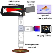
The PARISS imaging spectroscopy system is modular and designed for laboratory-based spectral and hyperspectral analysis. PARISS®: P rism A nd R eflector I maging S pectroscopy S ystem Basics The PARISS Prism: The Unique Curved Sides Dramatically Increases Sensitivity In order to increase instrument sensitivity, diffraction grating designs were discarded in favor of a prism-based system. The PARISS design was originally developed by The Aerospace Corporation for airborne remote sensing. After redesign, the PARISS imaging spectrometer emerged as an ideal instrument for use in any laboratory.The curved sides of the prism add optical power to deliver near aberration-free imaging, from 365 up to 1,000-nm.The net result is a highly significant improvement in signal to noise ratio (S/N) when compared to diffraction grating solutions. Enhanced sensitivity extends into the near IR where detectors and visible diffraction gratings are at their lowest efficiency. Spectrum cameras: Located at the focus of the imaging spectrometer, the camera records the sample spectra. Spectrum cameras are chosen as a function of anticipated signal strength.LightForm partners with camera manufacturers that supply the appropriate camera consistent with an application.Zoom light-collection optics: LightForm integrates lenses optimized for the application. For example, microscope objectives may be necessary for biopsy samples, while telephoto lenses would be used to image ponds polluted with deadly cyanobacteria. There is no one lens solution that fits all applications.LightForm specializes in developing zoom magnification optics that eliminate the need for multiple lenses or objectives. While zoom camera lenses are commonplace most, if not all de-magnify a field of view (FOV). PARISS zoom magnification optics accommodates objects that are less than one micron in size. Software: PARISS software is written in Python, with spectroscopy utilities that include: %reflection, absorption, %transmission, fluorescence, luminescence… Math functions include smoothing, background subtraction, division, multiplication, noise reduction… Spectral classification: Spectra present that can be associated with objects or conditions are assembled into classes. Selected classes can then be added to a “Reference Spectral Library.” Correlation functions enable the extent of a class to be controlled and optimized.Reference spectral libraries (RSL): RSLs consist of a collection of spectral classes consistent with a given application or sample type. RSLs can be created, added to, or edited. Spectral classes can be named and pseudo-colored Spectra recognition algorithms: Enables objects in new samples to be “recognized” by correlating their spectra with those in an RSL. A recognized object acquires the pseudo-color of the associated RSL component class. Imaging Spectroscopy Requires a Highly Efficient Imaging Spectrometer The PARISS imaging spectroscopy system is based on a prism imaging spectrometer:Unlike a diffraction grating that splits light into multiple diffraction orders, a prism delivers 90% of spectral intensity directly to the detector. This ensures the highest possible signal to noise ratio (S/N).
CATEGORY
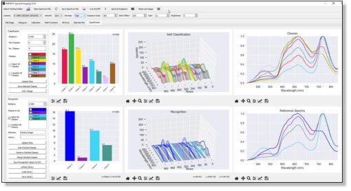
PARISS hyperspectral imaging software performs supervised and un-supervised spectral classification and creates reference spectral libraries.Signals buried in noise are revealed using spectral waveform cross-correlation (SWCCA) Pariss® Hyperspectral Imaging Field-scan-ning Software Workflow PARISS Spectral Imaging Spectroscopy Software Essentials Programing language: Python.Spectral classification: Unsupervised and supervised spectral classification. The user controls the number of revealed classes. Algorithms based on Spectral Waveform Cross-Correlation (SWCCA)Reference Spectral Libraries (RSL): A libraries of spectral classes that correlate with known objects or conditions. Classes can be added or removed, named and pseudo-colored by the user Spectral recognition: Correlates spectra presented by objects in the FOV with library spectra. The user selects the degree of correlation as a function of a user-selected minimum correlation coefficient (MCC) based on SWCCA.Spectroscopy functions: Math functions enable % Reflection, %Transmission, Fluorescence, Luminescence…Spectral plots: Single and multiple in 3D Camera control:Both the spectrum camera and observed image cameras Statistics:Display in a histogram or pie-chart format PARISS SWCCA Extracts Signal From Noise Even In Non-Linear Conditions Utilizes proprietary algorithms developed in house, based on Spectral Waveform Cross Correlation Analysis (SWCCA)Linearity independent SWCCA algorithms accommodate non-linear spectral mixing that normally occurs in biological samples.Highly tolerant of low S/N spectra: enables the generation of robust Reference Spectral Libraries (RSL) and correlation between sample spectra and RSL spectra.Reference Spectral Libraries that truly represent your samples.Enables accurate spectral segmentation.Standardized spectra Spectra acquired with PARISS can be standardized in absorption, % transmission/reflection.Publication ready spectra can be compared with spectra acquired on any other analytical instrument.Powerful topographical mapping: spectra from the FOV that “correlate” with RSL spectra can be pseudo-colored and “painted” onto a gray-scale image, with pixel-perfect accuracy. PARISS Hyperspectral Software Includes:Logical operators: “equals” and “not equals” at a user defined threshold. Controls risk of false positives or negatives.User created “real life” reference spectral libraries:Hyperspectral imaging software includes spectral topographical mapping: Map the location in a FOV of all or some target objects.SWCCA algorithms capture natural variations in spectral profile that indicate change such as: pH, ion-concentration, charge, and conformation.Measure change over time:PARISS can acquire spectra automatically over a user defined period, then play the results back as a movie.Observe raw spectra: all acquisitions can be exported to third party math or imaging programs.
CATEGORY
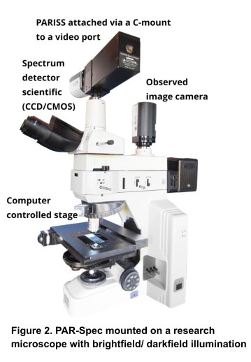
An Innovative Approach To Hyperspectral Microscopy Par- Spec acquires wavelength vs intensity spectra that correlate with CIE HSV ( Hue, Saturation, and Value) color-space.CIE coordinates are computed as a function of reflection spectra. Consequently, there is an intrinsic connection between reflection spectra and HSV coordinates. However, this relationship is one directional converting a HSV back to reflection spectra is not possible.Following the development of pseudo-colored, classified reference spectral and HSV libraries, spectral content of an entire FOV becomes a Hyperspectral Image with no further intervention. In Summary PAR-Spec Hyperspectral Imaging offers a comprehensive solution for laboratory-based hyperspectral imaging, meeting many of the goals of traditional hyperspectral imaging.PAR-Spec eliminates the need for tedious sample translation. User-selected pseudo-colors correlate with target objects or areas of interest, utilizing spectral libraries analogous to traditional hyperspectral imaging. Following the creation of a library an entire FOV is acquired in a single acquisition and analyzed based on %reflection/emission spectroscopy. Each spectrum is defined by hundreds of contiguous wavelength data points from 400 to 920 nm. Designed to work with objects as small as 1 micron 2, PAR-Spec is suitable for a wide range of applications, including particulates, microplastics, fibers, OLEDs, insects, forensic material, minerals, and vegetation. The PAR-Spec footprint is only 30 x 30 cm (excluding the lamp) makes it ideal for tight laboratory bench spaces. PAR-Spec is a system that includes the PARISS® imaging spectrometer mounted on a research-grade optical zoom microscope (ColSpec). Materials can be illuminated in reflection using either a 150W halogen lamp suitable for spectroscopic applications or a high-intensity (40,000 lumen) LED darkfield ring-light (see Figure 1).Additionally, PAR-Spec can be mounted on a research-grade microscope with dual ports, allowing two cameras to operate simultaneously PAR-Spec Pros•Once an spectral/HSV library has been established hyperspectral imaging future sample is a single acquisition to cover the entire field. •Covers a wide range of applications.•The height of a slice through the FOV is almost always less than the available pixels on a modern camera chip. The HSV acquisition covers the entire chip.•Spectral sampling target objects in a limited area of the FOV applies to the entire FOV.Cons•Less suited to applications that cover a limited spectral range. For example, some fluorescence experiments would be challenging. •HSV covers a very wide range of wavelengths, however some applications may present spectra outside the HSV range.•If a FOV is essentially monochromatic PAR-Spec will be ineffective PAR-Spec Eliminates Field-Scanning To Render a Hyperspectral Image In A Single Acquisition
CATEGORY
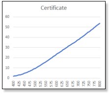
A NIST traceable light source to enable radiometric correction of a spectrometer output. Primarily for use with any microscope based spectrometer system. The SYLPH, NIST Certified, Radiometric Calibration Solution. The accepted way to normalize any spectrometer is to acquire a spectrum of NIST certified characteristics. For wavelength accuracy use an absolute light source such as the MIDL Hg/Ar emission lamp. For wavelength versus intensity use a NIST certified emission lamp such as the “SYLPH” radiometric calibration light source. The SYLPH radiometrically calibrated light source delivers a stable, NIST-certified spectrum of known characteristics. This provides a spectral microscope or spectral confocal system with a certified traceable “reference emission.” To standardize a system determine a correction curve by acquiring a spectrum of the SYLPH lamp and compare it to the spectrum described in the certificate. After applying the correction curve the spectrometer will reconstruct the spectrum of the SYLPH lamp and accurately reconstruct the spectrum of any other sample. Once this correction factor has been incorporated in the data processing software it should be applied to all future acquisitions. Bottom line: All radiometrically calibrated instruments will accurately reproduce the same reference spectrum. Once an instrument has been radiometrically corrected all spectra acquired on that instrument can be compared with those acquired on other radiometrically calibrated instrument. SYLPH: Radiometric Calibration System Specifications Source: Tungsten halogen Wavelength range: 360-2400 nm Warm-up time: 10 minutes at 23deg ambient Bulb power: 4.75 W (nom) Source lifetime: 10,000 hrs (typical) Calibrated for: Absolute irradiance (µW/cm2/nm) Stability of optical output: 0.15% peak-to-peak Drift of optical output: <0.3% per hour Operating temperature: 5 °C – 35 °C Humidity: 5-95% without condensation at 40 °C Power Consumption: up to 15 W “SYLPH™” Standard Yield Lamp for Photonic Calibration. NIST Certified radiometric power spectrum as a function of wavelength in relative µW/cm2/nm. Relative spectrometer spectrum of the NIST lamp before correction in arbitrary units. The SYLPH radiometric calibration system setup (not to scale. The illumination head is placed facing the microscope objective.
CATEGORY
