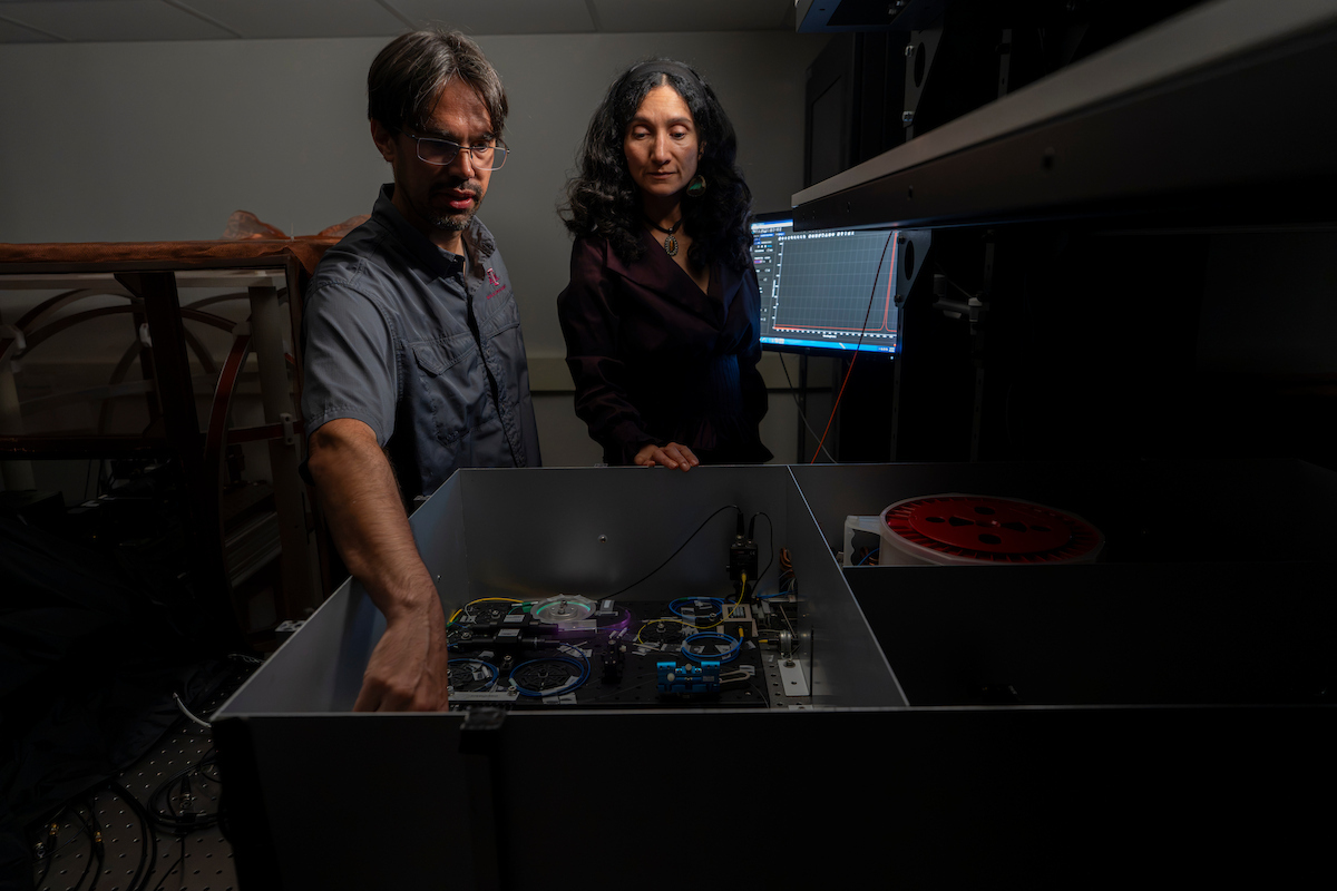
Researchers at Texas A&M University cracked a problem that has limited biological imaging for years: seeing fine cellular detail and looking deep inside living tissue at the same time.
The new microscopy technique resolves structures smaller than 100 nanometers, roughly a thousand times thinner than a human hair, without giving up the ability to image deep into intact samples.
The team, led by Aart Verhoef, assistant professor of biophotonics in the Department of Soil and Crop Sciences in the Texas A&M College of Agriculture and Life Sciences, and joint professor in the Department of Physics and Astronomy, published the findings in the Journal of Biomedical Optics.
Their method, called 2PE-ISM-SOFI, short for two-photon excited image scanning microscopy combined with super-resolution optical fluctuation imaging, layers two established techniques onto a standard two-photon microscope to sharpen its resolution without shrinking its reach.
The result is lateral resolution down to about 75 nanometers, roughly five times sharper than conventional two-photon microscopy, one of the most widely used tools for imaging living cells and tissue.
“This technique builds on two microscopy techniques that were limited by depth and detail, but this technique allows us to look deep into tissue and improves the resolution,” Verhoef said.
Bridging the gap between depth and detail
Two-photon microscopy earned its place in labs because it can peer deep into biological samples with minimal damage, a must for studying living tissue in fields ranging from neuroscience and developmental biology to soil-, animal- and plant-related sciences. But that depth has come with a trade-off: seeing fine detail has typically meant staying near a sample’s surface.
Super-resolution techniques such as Stimulated Emission Depletion, STED, and Photoactivated Localization Microscopy, PALM, which earned their developers the 2014 Nobel Prize in Chemistry, can resolve features far below 100 nanometers, but usually only close to the surface. The new method narrows the gap, holding on to two-photon imaging’s depth advantage while closing in on that sharper resolution.
Breakthrough opens door to deeper understanding at cellular levels
The team laid groundwork for the approach in a 2024 study in PRX Life. Together, the two studies pave a path for imaging tools that could reach further into plant and soil biology than current methods allow, capturing root structures, plant-microbe interactions or disease progression at the cellular level without trading away depth for detail.
Co-author Alma Fernandez Gonzalez, biophotonics researcher in the Department of Soil and Crop Sciences, said application of the technique in fields from neuroscience to plant and animal biology will take time.
“Our friends in the College of Medicine are looking at ways to look deeper and with more detail,” she said. “For this study, they cultured midbrain dopamine neurons and labeled the tubulin in the cells with quantum dots. We still have further to go to improve this technique, but we see a path to do that.”
Part of a statewide push in quantum science
The Texas A&M Division of Research has made quantum science an applied research priority, including quantum sensing tools such as ultra-sensitive bio-imaging. The Texas Legislature created the Texas Quantum Initiative in 2025 to guide quantum research, funding and manufacturing across the state. The microscopy project grew out of that push for cross-disciplinary research under the prioritized initiative.
Co-authors span the Department of Soil and Crop Sciences, Department of Physics and Astronomy, the Institute for Quantum Science and Engineering, the Department of Neuroscience and Experimental Therapeutics in the Texas A&M Naresh K. Vashisht College of Medicine, the Department of Biology, the Department of Biomedical Engineering and the Department of Biological and Agricultural Engineering.
Verhoef and several collaborators organized a panel on quantum sensing for biomedicine and agriculture at the second Texas Quantum Summit, held in August in Dallas.
“It is exciting for us to break a barrier that has been a challenge for ages and to open the door for new investigations and discoveries,” Verhoef said.
Reference
Breaking the 100-nm resolution barrier with multiphoton microscopy using image scanning microscopy and optical fluctuation imaging
IonQ and Carahsoft Technology Corp. announced a strategic partnership. Carahsoft will serve as IonQ's Public Sector distributor, enabling Government agencies to purchase IonQ technology through its reseller partners.
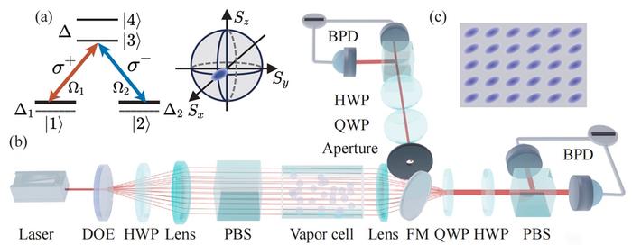
A 30-channel array of polarization-squeezed light has been generated in a single paraffin-coated rubidium vapor cell, with thermal atomic motion carrying a shared ground-state coherence between spatially separated beams. The collective coupling synchronizes the channels: a deliberately mismatched beam is pulled from a 0.79 dB to a 0.19 dB difference, and the array squeezes where a lone channel cannot.
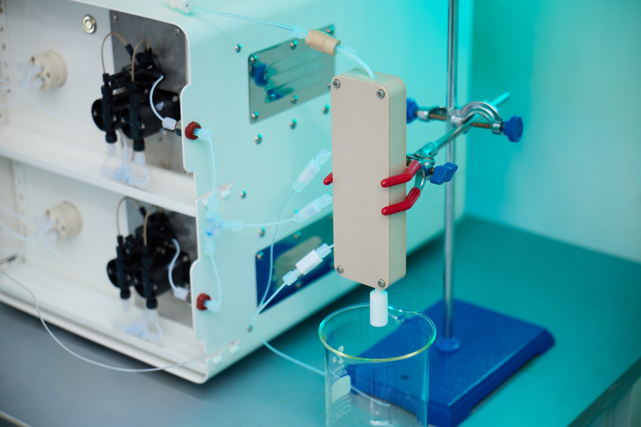
Accelerated Materials has launched the K1 Pro, an automated four-reagent microreactor for 1 kg/day nanoparticle synthesis. The company says processes developed on it move to its K10 and K100 systems with minimal reconfiguration.
This week in nanotech
$9M raised
1 funding round · 22 deals tracked
New products
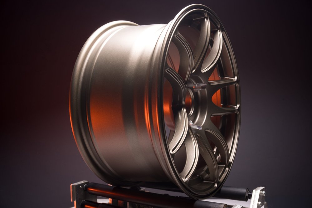 Ti-WCMBN Nanomaterialia Featured
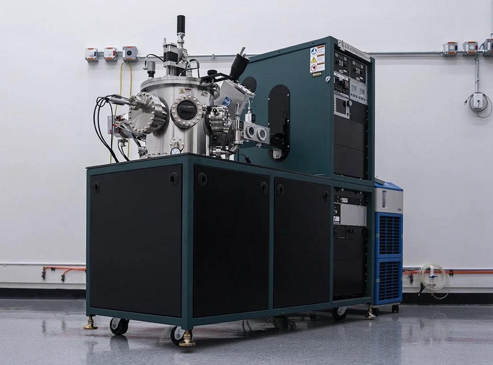 Orion Sputtering Deposition SystemsAJA International Featured
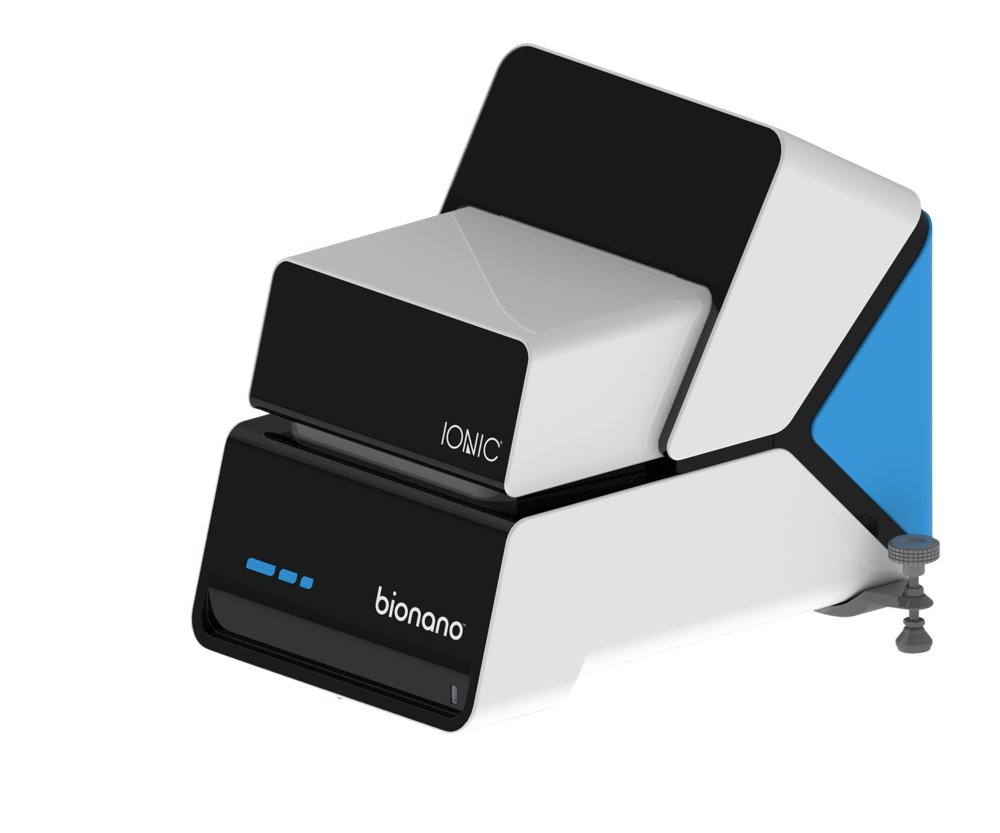 Ionic® Purification SystemBionano Genomics, Inc.
Market insights
Last 30 days: Quantum Technologies was the busiest vertical: 59 of 192 events (31%).
Money raised per month