Physicists at the University of Wisconsin–Madison directly measured, for the first time at nanometer resolution, the fluid-like flow of electrons in graphene. The results, which will appear in the journal Science on Feb. 17, have applications in developing new, low-resistance materials, where electrical transport would be more efficient.
Graphene, an atom-thick sheet of carbon arranged in a honeycomb pattern, is an especially pure electrical conductor, making it an ideal material to study electron flow with very low resistance. Here, researchers intentionally added impurities at known distances and found that electron flow changes from gas-like to fluid-like as temperatures rise.
“All conductive materials contain impurities and imperfections that block electron flow, which causes resistance. Historically, people have taken a low-resolution approach to identifying where resistance comes from,” says Zach Krebs, a physics graduate student at UW–Madison and co-lead author of the study. “In this study, we image how charge flows around an impurity and actually see how that impurity blocks current and causes resistance, which is something that hasn’t been done before to distinguish gas-like and fluid-like electron flow.
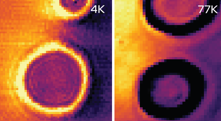
The researchers intentionally introduced obstacles in the graphene, spaced at controlled distances and then applied a current across the sheet. Using a technique called scanning tunneling potentiomentry (STP), they measured the voltage with nanometer resolution at all points on the graphene, producing a 2D map of the electron flow pattern.
No matter the obstacle spacing, the drop in voltage through the channel was much lower at higher temp (77 kelvins) vs lower temp (4 K), indicating lower resistance with more electrons passing through.
At temperatures near absolute zero, electrons in graphene behave like a gas: they diffuse in all directions and are more likely to hit obstacles than they are to interact with each other. Resistance is higher, and electron flow is relatively inefficient. At higher temperatures — 77 K, or minus 196 C — the fluid-like behavior of electron flow means they are interacting with each other more than they are hitting obstacles, flowing like water between two rocks in the middle of a stream. It is as if the electrons are communicating information about the obstacle to each other and diverting around the rocks.
“We did a quantitative analysis [of the voltage map] and found that at the higher temperature, the resistance is much lower in the channel. The electrons were flowing more freely and fluid-like,” Krebs says. “Graphene is so clean that we’re forcing the electrons to interact with each other before they interact with anything else, and that is crucial in getting them to behave like a fluid.”
Reference
Imaging the breaking of electrostatic dams in graphene for ballistic and viscous fluids
Zachary J. Krebs, Wyatt A. Behn, Songci Li, Keenan J. Smith, Kenji Watanabe, Takashi Taniguchi, Alex Levchenko, Victor W. Brar
https://www.science.org/doi/10.1126/science.abm6073
University of Wisconsin–Madison
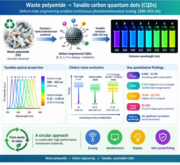
Waste polyamide-derived CQDs show continuous photoluminescence tuning from 308 to 552 nm through sequential defect-state engineering, with optical transition energies decreasing from 4.32 to 2.50 eV. @Christian Ebere Enyoh from Saitama University Carbon quantum dots (CQDs) are fluorescent carbon nanomaterials with potential applications in sensing, optoelectronics, displays, anti-counterfeiting, and environmental technologies. Their optical properties can be adjusted by modifying the carbon...
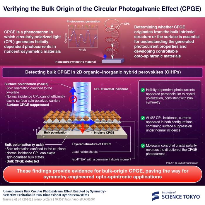
The researchers used circularly polarized light at normal incidence and found that helicity-dependent photocurrents appeared perpendicular to the crystal’s polarization but vanished when measured parallel to it, supporting a bulk origin of CPGE. @Institute of Science Tokyo The circular photogalvanic effect (CPGE), a phenomenon that generates helicity-dependent photocurrents in noncentrosymmetric materials, can originate purely from a crystal's internal structure without contribution from the...
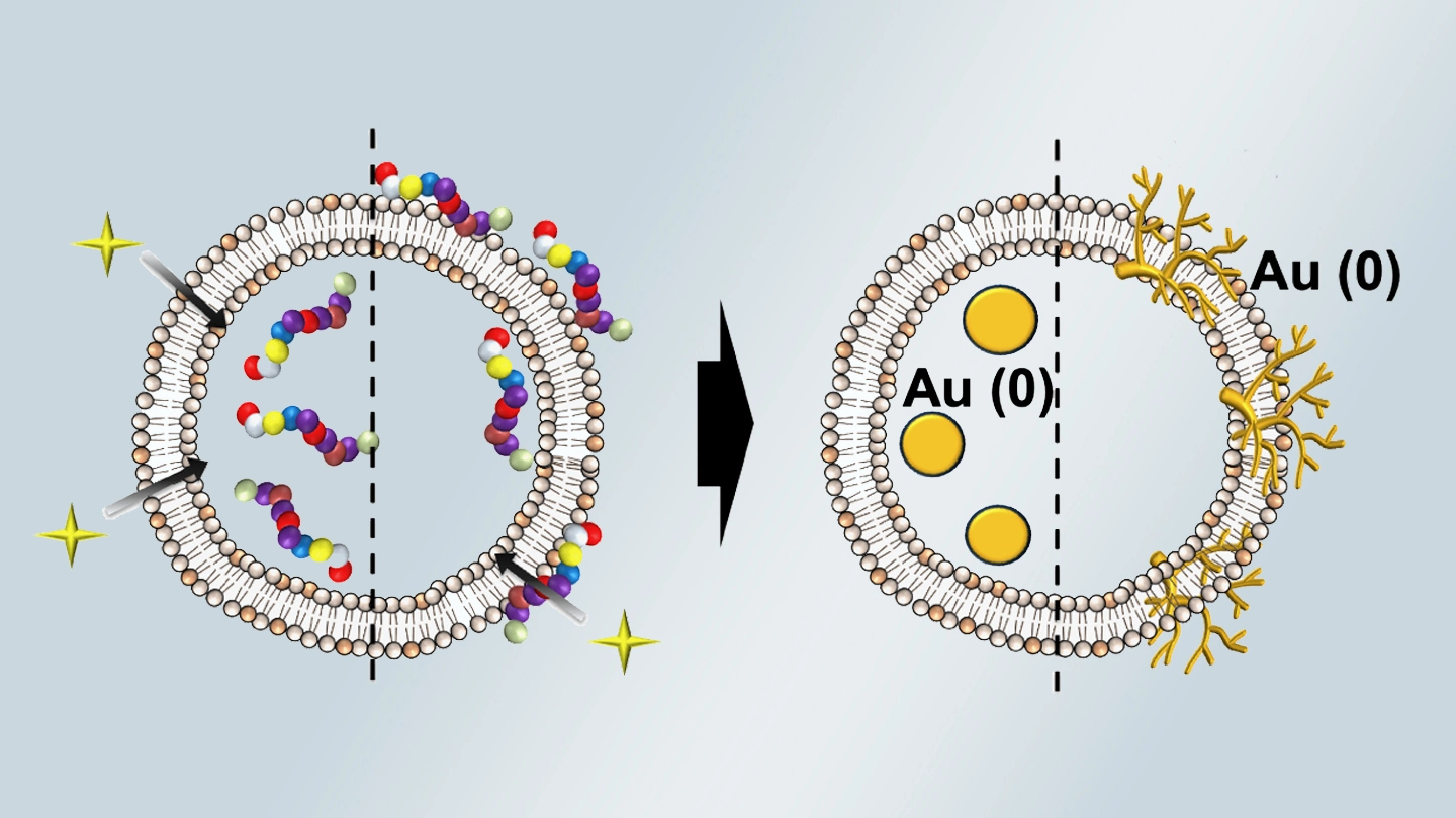
Summary of gold nanoparticle growth programming in liposome based on localization of biomineralization peptide The position of biomineralization peptides within liposomes can influence how gold nanoparticles grow, reports a research team from Institute of Science Tokyo. Peptides localized at the membrane interface promote branched structures, while those confined to the liposome interior favor spherical nanoparticles. The findings offer a new strategy for controlling nanoscale reaction...
