By Gatan
STEMx System Model 1020 A powerful tool that expands the utility of your Gatan camera to address 4D STEM diffraction studies. Advantages Introducing STEMx®, a powerful tool that adds 4D scanning transmission electron microscopy (STEM) diffraction capabilities to your existing Gatan camera (K3®IS, Metro® camera series, ClearView®IS, OneView®IS, Rio®16 IS, or Stela®*) and GIF Continuum®. 4D STEM is the capture of a full 2D diffraction pattern at each pixel position in a 2D STEM map. Powered by eaSI™ technology Ensures high-quality data for all users Unmatched 4D STEM across platforms Use the STEM spectrum imaging (SI) technique in DigitalMicrograph® to set up the experimental parameters, then start acquisition, and one diffraction pattern is automatically recorded for each STEM probe position via hardware synchronization through the STEMx box. Using the STEM SI technique, EELS, EDS, or CL signals are combined and collected simultaneously or consequentially from the same area of the specimen (acquisition mode dependent). Similarly, you can collect 5D STEM datasets (4D STEM + time) using the 4D STEM in-situ package. Such multimodal, linked data acquisition allows morphological and structural analysis via diffraction imaging and chemical and compositional analysis via spectroscopy and allows for a more conclusive analysis of your specimens. To ensure you get the most out of your information-rich 4D datasets, STEMx offers integrated analysis tools. This includes: - Virtual apertures - Strain mapping - Differential phase contrast - Orientation mapping and indexing Datasheet STEMx System A 4D STEM diffraction data cube collected using the Stela camera and STEMx system. Specimen: Au nanoparticles. Scan area: 176 x 218 pixels, Diffraction image size: 256 x 256 pixels. Dwell time: 5 ms. Color map showing grain orientations classified based on the angular position of the maximum diffraction peak per probe position using a DigitalMicrograph script.
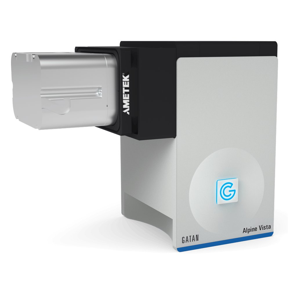
Models 1124, 1125. The world’s first direct detection cameras to democratize cryo-EM. Alpine®is the world’s first direct detection camera series to democratize cryo-electron microscopy (cryo-EM) imaging. By enabling the acquisition of high-resolution structures on 80 – 200 keV microscopes, the Alpine series allows routine cryo-EM research on more affordable, workhorse instruments and expands its usefulness to the larger research community. Two models available to match the field of view with your application needs: Alpine (1124) – Turns 80 – 200 keV screening microscopes into data-collection microscopes with single-electron counting capabilities. Delivers a detective quantum efficiency (DQE) up to 2.4-times the performance of scintillator-based cameras. Makes the 2D classification of small molecules possible while improving macromolecule reconstructions. Streamlines your workflow and detects failures early to produce better structures, faster. Real-time counting ensures there is no compromise between data collection speed and quality.
CATEGORY
Models 1124, 1125. The world’s first direct detection cameras to democratize cryo-EM. Alpine®is the world’s first direct detection camera series to democratize cryo-electron microscopy (cryo-EM) imaging. By enabling the acquisition of high-resolution structures on 80 – 200 keV microscopes, the Alpine series allows routine cryo-EM research on more affordable, workhorse instruments and expands its usefulness to the larger research community. Two models available to match the field of view with your application needs: Alpine Vista (1125) – Maximizes imaging throughput for your high-performance microscopes with a large FOV of 5,760 x 4,092 pixels. Delivers a detective quantum efficiency (DQE) up to 2.4-times the performance of scintillator-based cameras. Makes the 2D classification of small molecules possible while improving macromolecule reconstructions. Streamlines your workflow and detects failures early to produce better structures, faster. Real-time counting ensures there is no compromise between data collection speed and quality.
CATEGORY
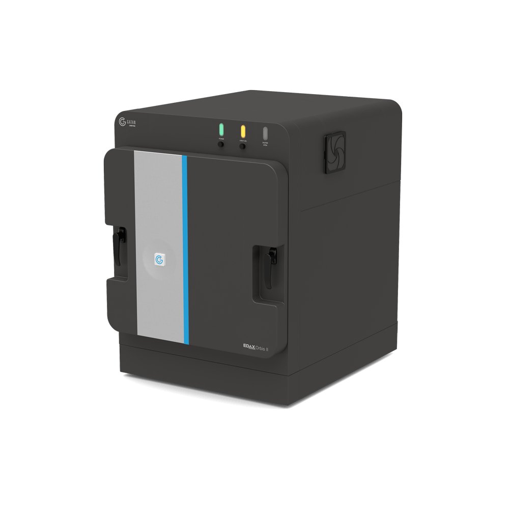
Model 1140 Delivers non-destructive, high-performance elemental analysis with unmatched flexibility and ease of use. The EDAX® Orbis™ II micro-x-ray fluorescence (micro-XRF) system delivers non-destructive, high-performance elemental analysis with unmatched flexibility and ease of use. Engineered for excellence across the full spectrum of micro-XRF applications, the Orbis II empowers users to analyze everything from the smallest particles to large, complex samples—quickly and accurately. Experience the next level of micro-XRF analysis with the EDAX Orbis II—where precision meets performance. - Custom x-ray window technology enables the detection of light elements - Proprietary coaxial light and x-ray optics precisely targets specific regions – eliminating x-ray interference from adjacent areas - Maximizes throughput by collecting spectra 2.5x faster and spectral maps 3x faster - Streamlines workflows with programmable autorun routines to boost productivity and ensure consistent results
CATEGORY
