By Photon etc
CIMA is a hyperspectral confocal system sensitive between 400 and 1700 nm. Spectral resolution can be as low as 0.2 nm in the visible range, and 0.6 nm in the infrared. It pairs a galvanometer head and one of the fastest and most sensitive cameras on the market to yield an acquisition rate above 300 spectra per second. CIMA provides three acquisition modes: confocal hyperspectral imaging, multispectral fluorescence imaging, and emission spectroscopy of a sample in a cuvette. CIMA APPLICATIONS OVERVIEW: - Simultaneously study the spatial distribution and spectral properties of complex nanomaterials such as upconverting nanoparticles and quantum dots. - Perform in vitro or in vivo high resolution hyperspectral microscopy. - Develop new sensors based on environment dependent spectral shifting of their fluorescence signal. TECHNICAL SPECIFICATIONS Spectral range 400 - 1700 nm Spectral resolution VIS < 0.2 nm IR < 0.6 nm Custom resolution upon request Spatial resolution Diffraction limited Cameras Back-illuminated CCD or EMCCD; InGaAs linear array Excitation wavelength 980 nm (Other wavelengths available) 3 laser input ports UHP mercury lamp 130W Microscope Scientific grade, inverted Objectives 20X, 40X, 60X; 50X, 100X Other magnifications available upon request Wavelength absolute accuracy 0.25 nm Maximum scanning speed > 300 spectra/s ~ 100 spectra/s Motorized XYZ stage 120 x 75 x 0.15 mm Preprocessing Spatial filtering, statistical tools, spectrum extraction, data normalization, spectral calibration Hyperspectral data format HDF5, FITS Software PC (Windows10 - 64-bits) with PHySpec™ control and analysis software (computer included) Power requirement 120 VAC / 12A / 60Hz; 230 VAC / 12A / 50Hz CONFOCAL MODE Scanning region 300 μm x 300 μm (20x objective); 100 μm x 100 μm (60x objective) Pinhole diameter 30 to 200 μm CUVETTE MODE Cuvette size Standard 10 mm x 10 mm (3.5 mL) FLUORESCENCE MODE Camera Megapixel colour camera for fluorescence and sample visualization Illumination lamp UHP mercury lamp 130W Epifluorescence filter cubes Up to 6 different filter cubes
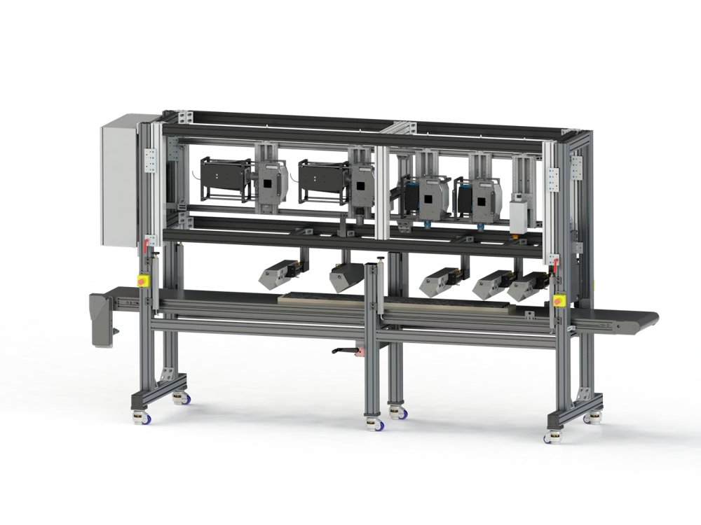
Hyperspectral Core Scanner The nCore is a multi-sensor scanning platform designed for drill core analysis. Whether deployed directly on-site or in a laboratory setting, the nCore offers unmatched versatility with modular configuration options tailored to the mining industry's needs. With the capability to integrate up to seven optical sensors, the nCore delivers an unparalleled map of the mineralogy landscape. Powered by Photon etc.'s proprietary hyperspectral imagers, it ensures precise and reliable performance for advanced mineral analysis, making it an indispensable tool for modern mining operations.
CATEGORY

Lecteur de plaques dédié à l’infrarouge à ondes courtes Lecteurs de plaques SWIR Photon etc. fait progresser le criblage à haut débit avec ClaIR™, un lecteur de plaques dédié à l’infrarouge à ondes courtes (SWIR, 900–1700 nm) et optimisé pour les nanocapteurs optiques et les nanomatériaux de prochaine génération. En opérant là où les matrices biologiques sont hautement transparentes, cette plateforme offre la sensibilité exceptionnelle requise pour cribler les capteurs à nanotubes de carbone à paroi simple (SWCNT) et suivre les éléments de reconnaissance moléculaire. ClaIR™ permet un balayage de zone automatisé par puits pour valider efficacement les nanocapteurs optiques dédiés aux cytokines inflammatoires, cartographier la localisation intracellulaire des capteurs en cytologie cancéreuse et accélérer la détection des biomarqueurs. ClaIR™ ClaIR est un lecteur de microplaques dans le NIR-II. Il enregistre la fluorescence de 900... Recherche sur les nanotubes de carbone
CATEGORY
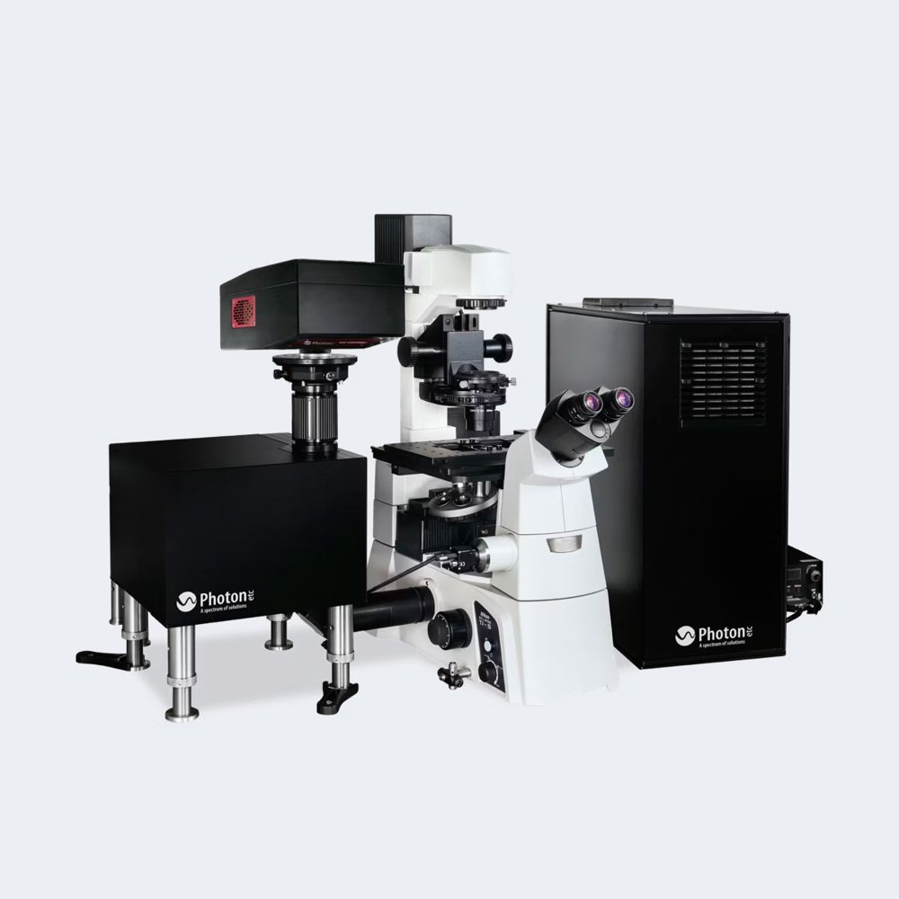
IMA is a hyperspectral microscope delivering spectral and spatial information from 400 nm to 1620... High-resolution spatial and spectral characterization at the microscopic scale
CATEGORY
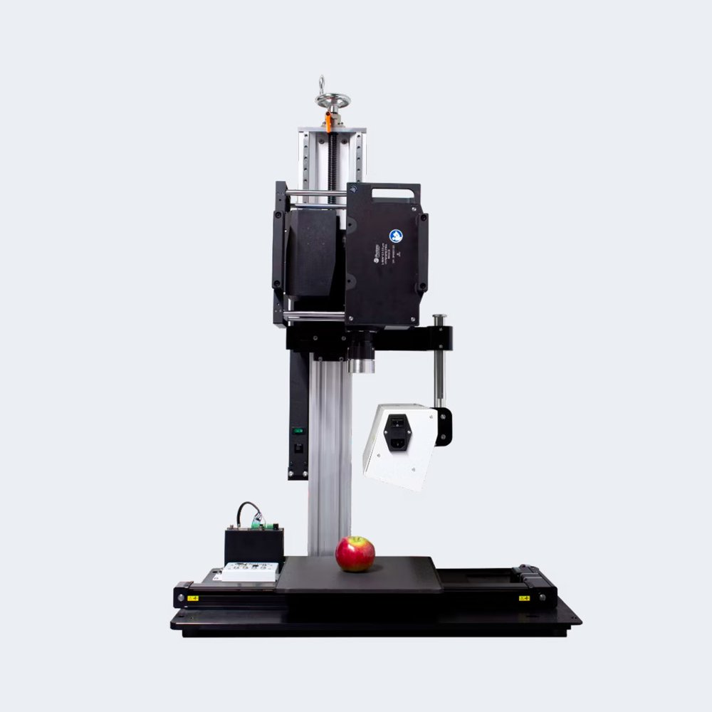
L-EOS™ is an infrared push broom hyperspectral SWIR camera 0.9 to 2.5 µm. In contrast to traditional imaging techniques that capture images in only three spectral bands (red, green, and blue) per pixel, the L-EOS, a hyperspectral imaging system, captures hundreds of spectral bands stacked in each pixel, forming a hyperspectral data cube. This unique capability allows for highly detailed and comprehensive spectral analysis of the imaged objects. However, it’s worth noting that the L-EOS requires either motion from the object or the camera itself to acquire the data cube, as it creates an image by scanning one row at the time. Hyperspectral data cube provides more information than single band imaging, making it suitable for material sorting and in-depth analysis.
CATEGORY
