15 October 2026. An experimental nanoparticle developed by Cedars-Sinai Health Sciences University investigators has been selected for evaluation by the Nanotechnology Characterization Laboratory (NCL). The program is funded by the National Cancer Institute and is designed to help promising nanotechnologies advance toward clinical trials.
The therapeutic nanoparticle, called HerDox2.0, was developed by Lali Medina-Kauwe, PhD, professor of Biomedical Sciences at Cedars-Sinai and associate director of Basic Research at Cedars-Sinai Cancer. The particle connects with a protein called HER3, which helps it cross the blood-brain barrier and target and penetrate tumor cells.
"Acceptance into the elite Nanotechnology Characterization Laboratory program will provide thorough testing of HerDox2.0 by top experts in the field, moving us toward our goal of submitting it to the Food and Drug Administration as an Investigational New Drug," Medina-Kauwe said.
The NCL analysis will determine the physical and chemical properties of the formulation, evaluate the particle's compatibility with and stability in human blood, assess potential immune responses, and evaluate general toxicity. Testing will also evaluate whether the particle can be manufactured consistently and in larger-scale batches for clinical use.
The Nanotechnology Characterization Laboratory, operated at the Frederick National Laboratory for Cancer Research, was founded in 2004 in collaboration with the Food and Drug Administration and the National Institute of Standards and Technology. To date, the laboratory has tested more than 1,000 nanomaterials and worked with more than 250 academic researchers and companies.
Source: Cedars-Sinai news
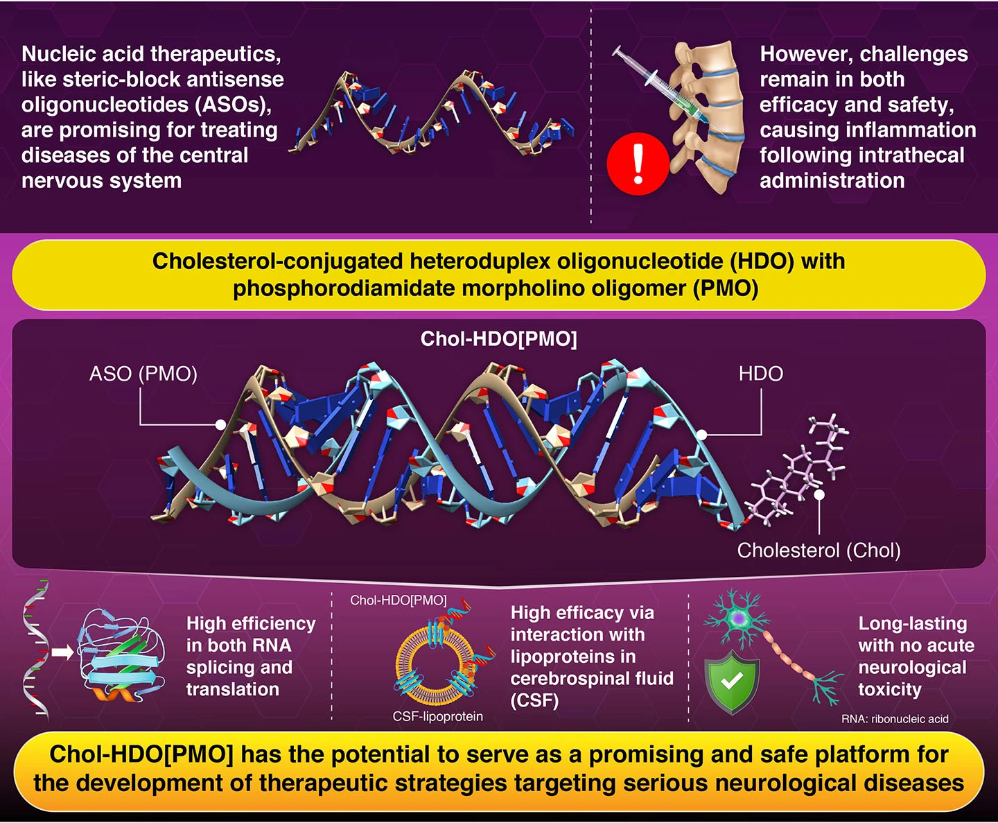
Researchers at the Institute of Science Tokyo have developed Chol-HDO[PMO], a cholesterol-conjugated heteroduplex oligonucleotide pairing a phosphorodiamidate morpholino oligomer with complementary RNA. Delivered into cerebrospinal fluid, it silenced or modulated target genes in mice with up to 16-fold greater potency than conventional antisense designs, and the effect was long lasting. It avoided acute neurotoxicity, weight loss and immune reactions.
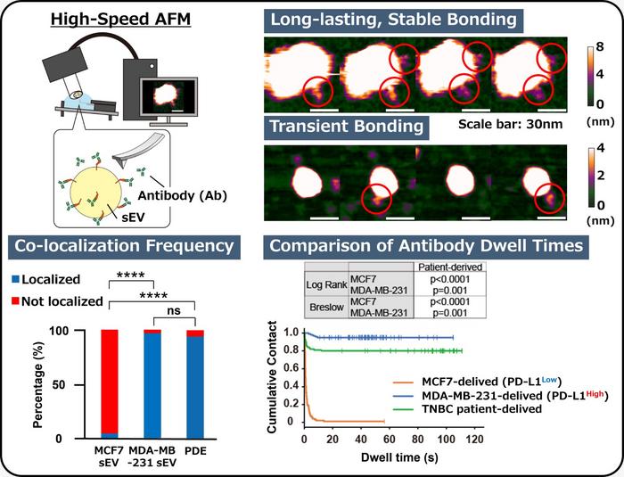
High-speed atomic force microscopy has revealed that the amount of PD-L1 on cancer-derived extracellular vesicles does not indicate how accessible the protein is to antibodies. Researchers at Kanazawa University imaged individual vesicles from two breast cancer cell lines in liquid, finding that over 80 percent of high-PD-L1 vesicles held antibodies persistently, while most low-PD-L1 vesicles showed no stable binding.
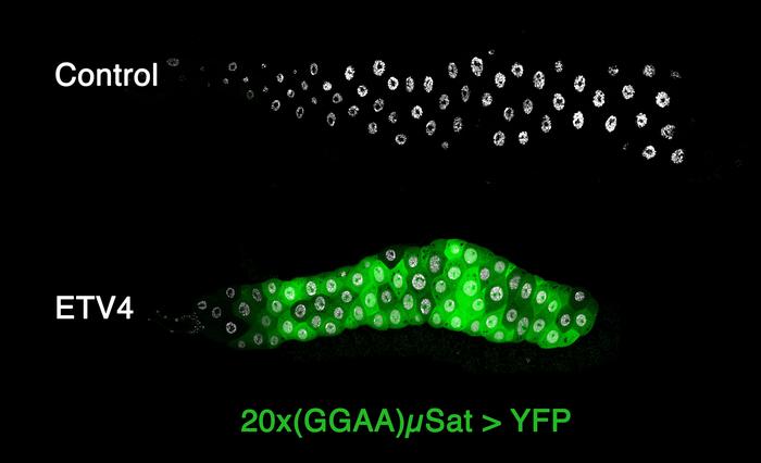
A study in Drosophila reveals that ETV4, an ETS-family protein implicated in prostate cancer, activates normally silenced GGAA microsatellite DNA regions by cooperating with an independent FET-family protein, Cabeza. The finding shows this activation mechanism does not require a fusion protein and is conserved across species, since human EWSR1 restores activity and ETV6 blocks it. The work broadens understanding of how repetitive DNA becomes regulatory in tumours beyond Ewing sarcoma.