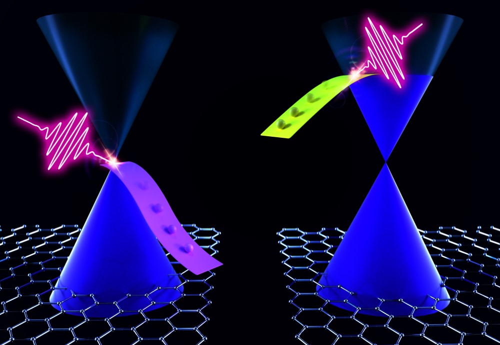
Graphene is the thinnest material ever produced, with the thickness of a single atomic layer, thinner than a billionth of a meter, it is able to efficiently absorb light from the visible to the infrared through the photoexcitation of its charge carriers. After light absorption, its photoexcited charge carriers cool down to the initial equilibrium state in a few picoseconds, corresponding to a millionth of a millionth of a second. The remarkable speed of this relaxation process makes graphene particularly promising for a number of technological applications, including light detectors, sources and modulators.
A recent study published in ACS Nano has shown that the relaxation time of graphene charge carriers can be significantly modified by applying an external electrical field. The research was conceived within an international collaboration between the CNR-IFN (Milano, Italy), Politecnico di Milano (Italy), the University of Pisa (Italy), the Cambridge Graphene Centre (UK) and the Catalan Institute of Nanoscience and Nanotechnology (ICN2, Barcelona, Spain).
“The change in the relaxation time of charge carriers in graphene that we have observed, demonstrates an unprecedented level of control on the optical response of a crystal and allows to obtain a large variety of behaviors using a single material,” says Dr Eva Pogna, researched from CNR-IFN, first author of the work. This study paves the way to the development of devices that exploit the control of the relaxation time of charge carriers to support novel functionalities. For example, if graphene is used as saturable absorber in a laser cavity to generate ultrashort light pulses, by changing the relaxation time of the charge carriers, we can control the duration of the output pulses.
“The specific device that we have used to study graphene, proved to be crucial to observe the strong tunability of its optical properties with the external electric field, allowing to change the number of charge carriers over a broad range by exploiting ionic liquid gating, which is a state-of-the-art technology introduced to study superconductors,” explains Prof. Andrea Ferrari, director of the Graphene Center in Cambridge.
The graphene-based device has been studied by ultrafast spectroscopy, which allowed to monitor the variation of the relaxation time of the charge carriers. “This work represents the latest step of a long-standing research collaboration devoted to the study of the ultrafast carrier dynamics in graphene, aimed at exploring the great potential of this fascinating material,” added Dr Klaas-Jan Tielrooij, leader of the Ultrafast Dynamics in Nanoscale Systems Group at ICN2.
Reference
Electrically Tunable Nonequilibrium Optical Response of Graphene
Eva A. A. Pogna, Andrea Tomadin, Osman Balci, Giancarlo Soavi, Ioannis Paradisanos, Michele Guizzardi, Paolo Pedrinazzi, Sandro Mignuzzi, Klaas-Jan Tielrooij, Marco Polini, Andrea C. Ferrari, and Giulio Cerullo
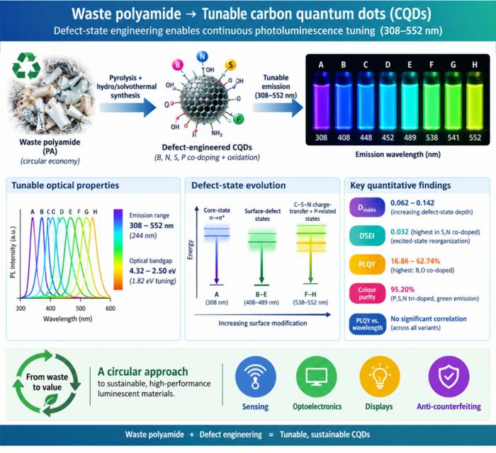
Waste polyamide-derived CQDs show continuous photoluminescence tuning from 308 to 552 nm through sequential defect-state engineering, with optical transition energies decreasing from 4.32 to 2.50 eV. @Christian Ebere Enyoh from Saitama University Carbon quantum dots (CQDs) are fluorescent carbon nanomaterials with potential applications in sensing, optoelectronics, displays, anti-counterfeiting, and environmental technologies. Their optical properties can be adjusted by modifying the carbon...
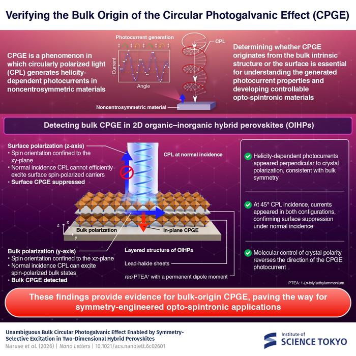
The researchers used circularly polarized light at normal incidence and found that helicity-dependent photocurrents appeared perpendicular to the crystal’s polarization but vanished when measured parallel to it, supporting a bulk origin of CPGE. @Institute of Science Tokyo The circular photogalvanic effect (CPGE), a phenomenon that generates helicity-dependent photocurrents in noncentrosymmetric materials, can originate purely from a crystal's internal structure without contribution from the...
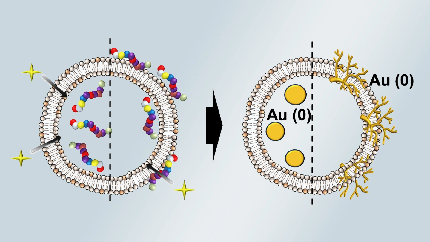
Summary of gold nanoparticle growth programming in liposome based on localization of biomineralization peptide The position of biomineralization peptides within liposomes can influence how gold nanoparticles grow, reports a research team from Institute of Science Tokyo. Peptides localized at the membrane interface promote branched structures, while those confined to the liposome interior favor spherical nanoparticles. The findings offer a new strategy for controlling nanoscale reaction...