One of the enigmas of life is emergence, when the whole becomes more than its parts. Flocks of birds can instantly change direction when a predator appears, guided not by a lead bird but by a collective intelligence that no single bird can possess on its own.
Multitudes of molecules skitter chaotically in a cell, but certain ones find each other, interact, and give rise to sophisticated cellular structures and functions that could not have been predicted by studying the molecules alone.
Understanding how emergent properties arise in cells – in this case, how liquid droplets called condensates spontaneously form from rapidly moving molecules -- “is a really hard problem, especially if we want to connect molecular properties to condensate properties,” says Michael Rosen, a Whitman Scientist at Marine Biological Laboratory (MBL) from University of Texas Southwestern Medical Center.
But this week in Science, Rosen and 10 other members of a large Whitman Center collaboration, the Chromatin Consortium, propose a long-awaited model for how the properties of a condensate can emerge from the properties of the individual molecules that compose it.
Their discoveries focus on chromatin, the densely compacted material in chromosomes that consists of our genetic information – long strands of DNA -- wound tightly around proteins. (See the Science commentary, "A Vision of Chromosome Organization.")
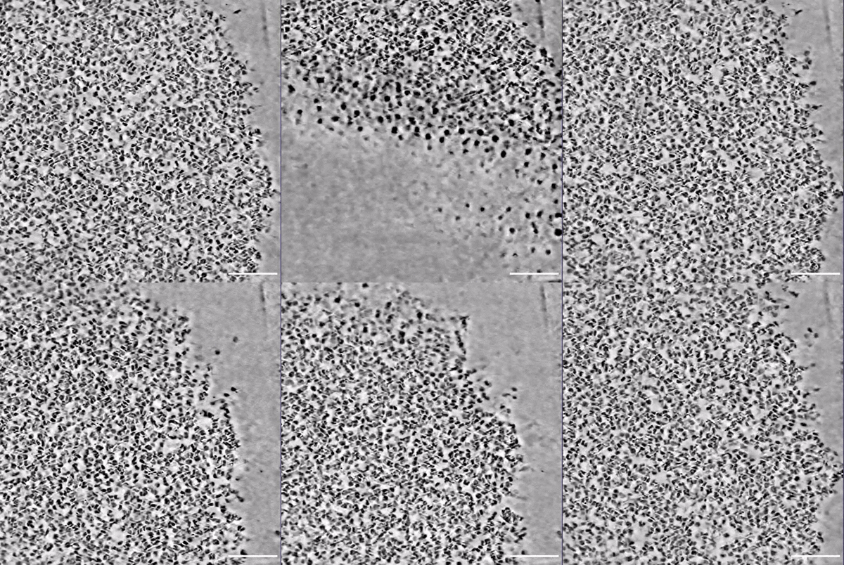
In the study, the scientists combined two powerful technologies – imaging (cryoET) to visualize the basic units of chromatin (the nucleosomes) and advanced computer simulations to explore how those units link together to form condensates – “to give us a view of a condensate that we just haven’t had before, in terms of the detail and the level of understanding that’s been provided,” says Rosen.
In chromatin, nucleosomes are linked together like beads on a string, connected by stretches of linker DNA. The length of the linker DNA, it turns out, is very important in determining the structure of successive nucleosomes on the string, and those nucleosome structures ultimately define how the molecules bind together to make the condensate.
Beyond chromatin, the new work provides a blueprint for studying and understanding the organization and function of many types of condensates. These membrane-less blobs carry out many important functions all over the cell — from regulating gene expression to responding to stress.
Understanding how these droplet-like structures form and function can help researchers understand what happens when condensation goes awry, a potential contributing factor for different diseases — from neurodegenerative conditions to cancer.
“By doing this research, we will better understand how abnormal condensation could lead to different diseases and, potentially, that could help us develop a new generation of therapeutics,” says Huabin Zhou, a postdoctoral scientist in the Rosen Lab and the lead author of the new research.
The Essential Role of the MBL
Ever since condensates were observed forming in live cells during the 2008 MBL Physiology course, “one of the long-standing goals in the community has been to connect the way molecules behave to the way condensates behave,” Rosen says.
In 2012, in an important paper in Nature, the Rosen lab proposed a biochemical mechanism for condensate formation in vitro. And seven years later, his lab was the first to formally recognize the capacity of chromatin to form condensates.
Yet visualizing the actual transition from a collection of molecules to a liquid droplet remained elusive to understanding.
“It’s a transition of scales,” Rosen says. “Molecules are on nanometer scales and condensates are on micron scales, and condensates have properties that are unique to their scale, like viscosity. You can’t talk about the viscosity of a molecule. Viscosity is something that arises when you get thousands or millions of molecules together to form a liquid.”
The Chromatin Consortium has been convening at MBL for the past three summers. The cryo-ET (imaging) data, which they obtained at HHMI Janelia and brought to Woods Hole, showed them where the units of chromatin (the nucleosomes) were located. At MBL, they wrestled to scale that data to higher resolution, using computer simulations led by Rosana Collepardo-Guevara of University of Cambridge, to understand exactly how the nucleosomes stick together to produce the condensate.
“To uncover the molecular details buried within the cryo-ET images, we had to design a new computer model (a coarse-grained model ) that could both scale up to condensates and faithfully capture the underlying chemistry in the nucleosomes,” Collepardo-Guevara says.
“Coarse-grained models always simplify reality; as the saying goes, all models are wrong, but some are useful. What made ours useful was the MBL,” Collepardo-Guevara says. “Being together for weeks over three summers, poring over cryo-ET maps and other experimental data, testing simulations, and debating what the data were really telling us, created the insight this problem needed. At the MBL, our simulation strategy grew from an idea into a powerful method that my group could not have built alone. The MBL brings a level of focus and collective interdisciplinary thinking that enables breakthroughs.”
Reference
Multiscale structure of chromatin condensates explains phase separation and material properties
Huabin Zhou, Jan Huertas, M. Julia Maristany, Kieran Russell, June Ho Hwang, Run-Wen Yao, Nirnay Samanta, Joshua Hutchings, Ramya Billur, Momoko Shiozaki, Xiaowei Zhao, Lynda K. Doolittle, Bryan A. Gibson, Andrea Soranno, Margot Riggi, Jorge R. Espinosa, Zhiheng Yu, Elizabeth Villa , Rosana Collepardo-Guevara, Michael K. Rosen
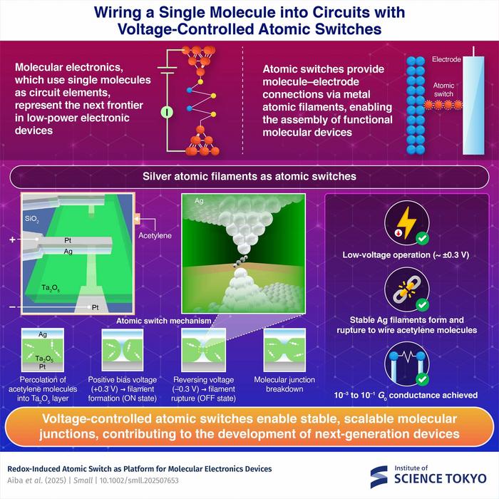
Silver-based atomic switches that create stable electrical connections between individual molecules and electrodes have been developed by researchers from Japan, addressing a key challenge in wiring molecular electronics. The switch operates by forming and breaking silver atomic filaments when a voltage is applied and reversed, corresponding to the “on” and “off” states. This method enables the scalable integration of molecular components, paving the way for ultra-compact and energy-efficient...
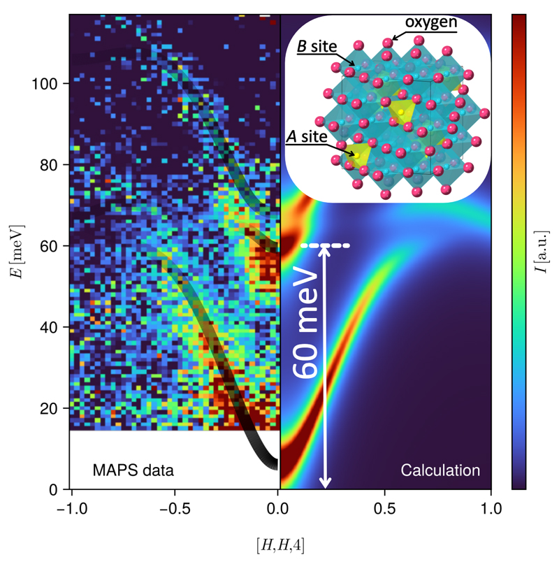
By measuring the spin dynamics over a broad energy range with neutron spectroscopy on a single crystal, the team identified a large band splitting of about 60 millielectronvolts (meV) between two magnon branches, a 3 meV anisotropy gap in the lower branch, and an avoided crossing near 75 meV in the upper branch. The researchers were then able to reproduce these important features quantitatively using theoretical calculations based on spin-wave theory.
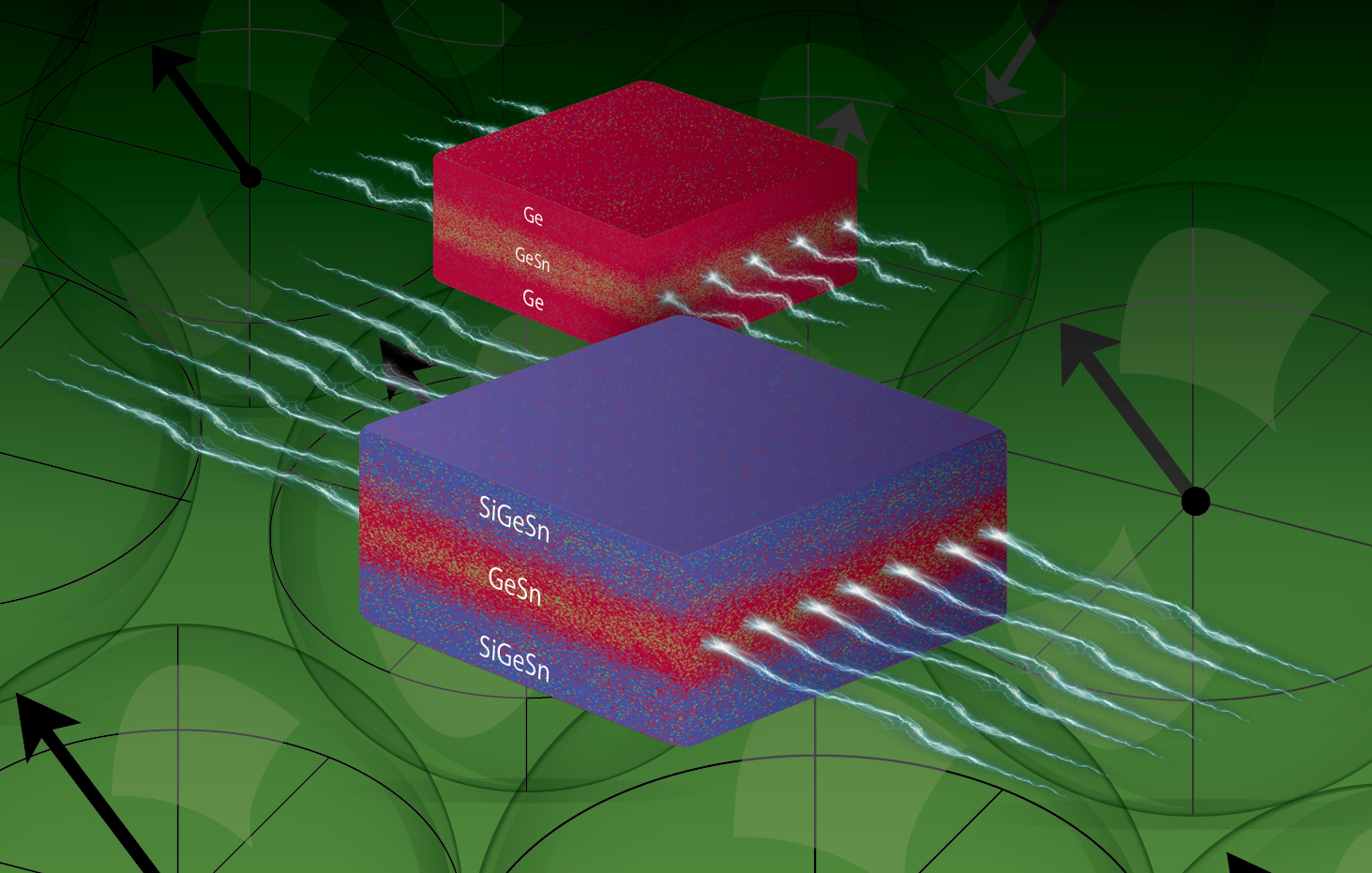
To visualize a quantum well, imagine a marble rolling in a groove between two raised edges. The marble can only move back and forth. A quantum well controls electrical current in a similar way, confining it in an ultrathin layer of material. This confinement improves how quickly you can encode information in light. The new paper shows how to make these wells work even better, whether for quicker downloads and smoother online experiences or for better qubits and more efficient transmission of...
This week in nanotech
$24.4M raised
5 funding rounds · 33 deals tracked
New products
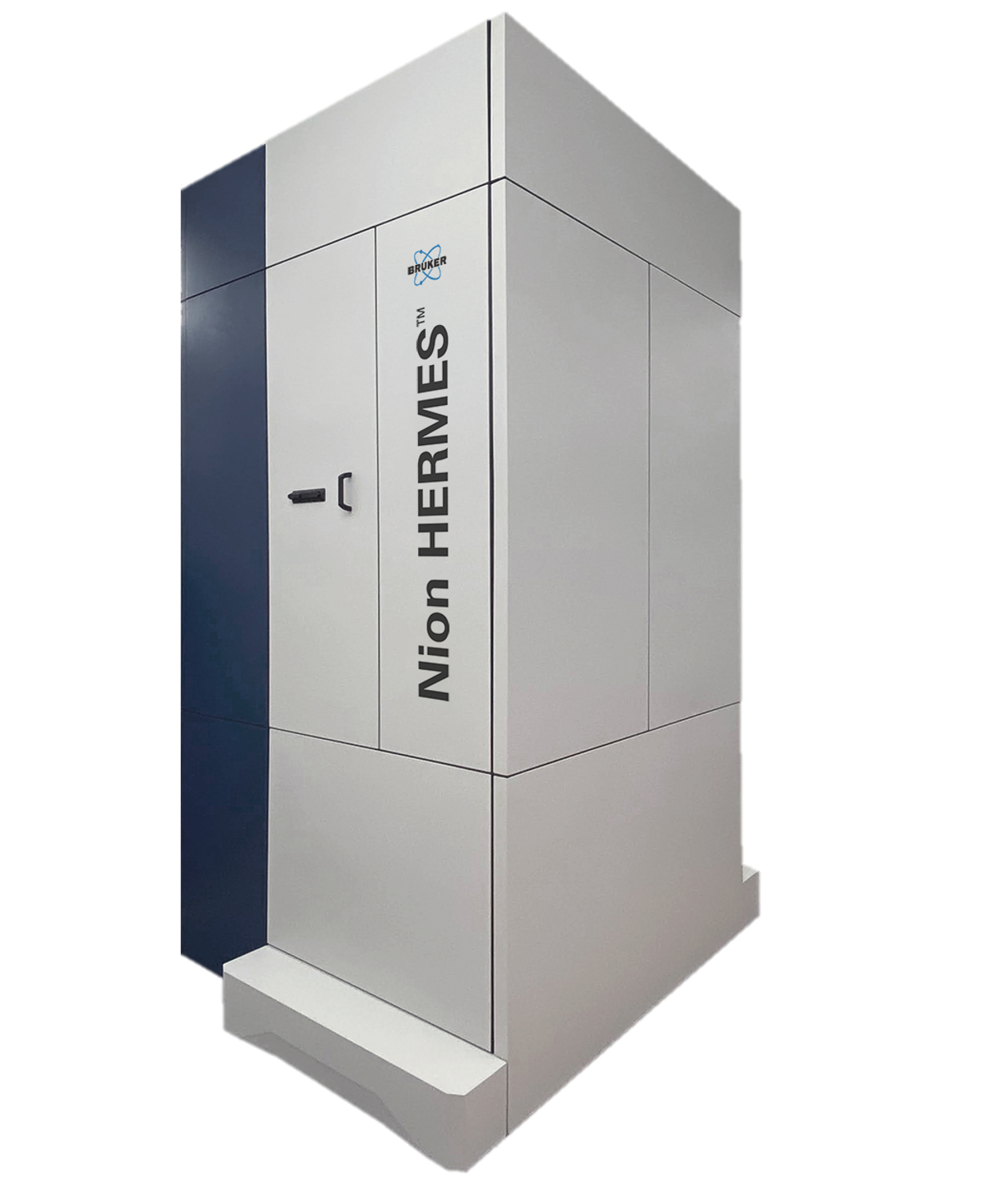 Nion-compatible In Situ STEM Holders for Electrical BiasingHummingbird Scientific Featured
 Ti-WCMBN Nanomaterialia Featured
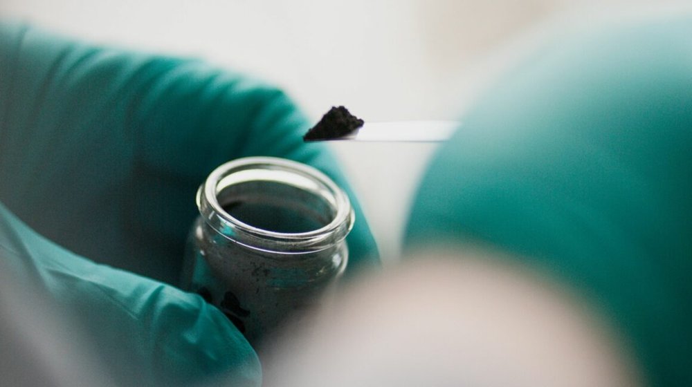 Talphene™ Graphene AdditivesTalga Group
Market insights
Last 30 days: Quantum Technologies drew the most money: $3.17B.
Money raised per month