
Imagine standing by a lake and throwing a stone into the water. Waves spread out in circular patterns and can reflect at obstacles and boundaries. Researchers at the University of Regensburg, in collaboration with colleagues from Milan and Pisa, have recreated this everyday phenomenon in a fascinating miniature world: They observed the propagation of waves – not on water but in an "electron sea" – using one of the fastest slow-motion cameras on the nanoscale.
Such electron seas are typically found on the surfaces of metals or materials with metallic properties. In this case, the material was graphene – a so-called two-dimensional material composed of a single layer of carbon atoms. Instead of a stone, the scientists used laser pulses, focusing them on a sharp metallic tip positioned just above the material's surface. ”The light sets the electrons in the tip in motion,” explains Simon Anglhuber from the Institute of Experimental and Applied Physics of the UR. “The resulting oscillations exert a force on the electrons in graphene. This generates a circular electron density wave that propagates through the graphene beneath the tip. The wave can reflect off the edges of the sample and travel back to the tip. These reflections can then be measured optically by reversing the previous process and converting the electron wave back into light. By precisely moving the tip over the sample, the researchers could record a film showing the wave's oscillation at various locations over time.
High-precision analysis of wave motion
The new technique allows for the direct observation of electron wave propagation in both space and time. This was achieved with a resolution on the nanometer scale – relevant for modern semiconductor technologies (1 nm = 10-9 m) – and a temporal resolution in the femtosecond range. In terms of temporal resolution, the method can be compared to an ultra-fast slow-motion camera with a frame rate of over 10 trillion frames per second (>10¹³ fps). The result is a highly precise analysis of wave motion, including its speed, damping, and frequency, without requiring complex computational transformations. Notably, the researchers observed a distinction between the propagation of the wave's center of mass and the propagation of individual wave peaks and troughs. By precisely measuring these two speeds, it is possible to infer the properties of the material through which the waves are propagating.
In their experiments, the researchers compared graphene samples produced by different methods and found significant differences in wave propagation, which were linked to variations in sample quality. These findings are expected to contribute to the development of better samples for use in optoelectronic devices, such as highly sensitive light sensors. Remarkably, the method also works for heavily damped electron waves in the so-called terahertz and mid-infrared range – a spectral region between our 5G network and visible light that has been difficult to access so far.
Ultrafast control of surface waves
As a final step, the researchers used another laser pulse to deliberately perturb the electron sea in the graphene sample while the electron wave was propagating. By including the second laser pulse, they were able to selectively weaken the wave. This not only allows for observation of the waves and insights into the material in its static form but also enables control and ultrafast alteration of material properties. This direct control of electron density waves could be a key step toward developing new electronic components with clock speeds more than a thousand times faster than current electronics.
Reference
Spacetime Imaging of Group and Phase Velocities of Terahertz Surface Plasmon Polaritons in Graphene
Simon Anglhuber, Martin Zizlsperger, Eva A. A. Pogna, Yaroslav A. Gerasimenko,
Anastasios D. Koulouklidis, Imke Gronwald, Svenja Nerreter, Leonardo Viti, Miriam S. Vitiello, Rupert Huber & Markus A. Huber
https://pubs.acs.org/doi/10.1021/acs.nanolett.4c04615
Universität Regensburg
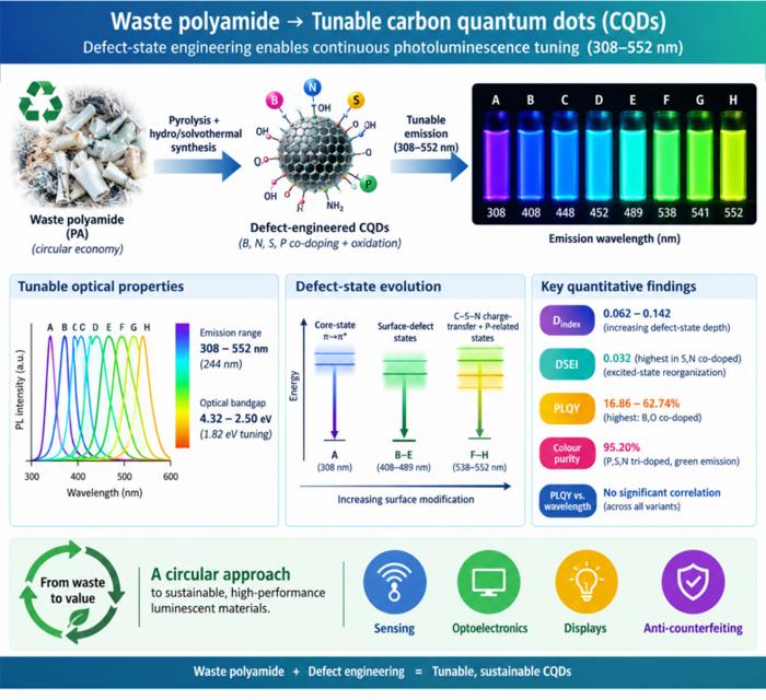
Waste polyamide-derived CQDs show continuous photoluminescence tuning from 308 to 552 nm through sequential defect-state engineering, with optical transition energies decreasing from 4.32 to 2.50 eV. @Christian Ebere Enyoh from Saitama University Carbon quantum dots (CQDs) are fluorescent carbon nanomaterials with potential applications in sensing, optoelectronics, displays, anti-counterfeiting, and environmental technologies. Their optical properties can be adjusted by modifying the carbon...
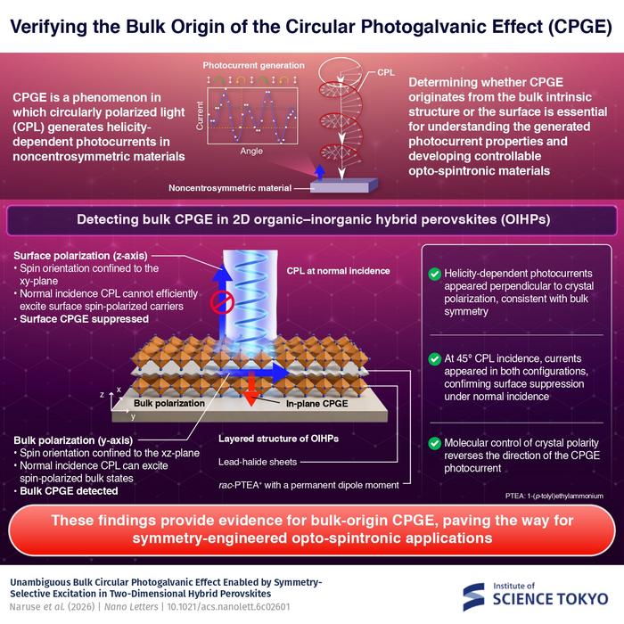
The researchers used circularly polarized light at normal incidence and found that helicity-dependent photocurrents appeared perpendicular to the crystal’s polarization but vanished when measured parallel to it, supporting a bulk origin of CPGE. @Institute of Science Tokyo The circular photogalvanic effect (CPGE), a phenomenon that generates helicity-dependent photocurrents in noncentrosymmetric materials, can originate purely from a crystal's internal structure without contribution from the...
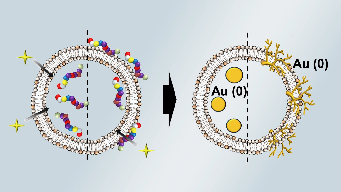
Summary of gold nanoparticle growth programming in liposome based on localization of biomineralization peptide The position of biomineralization peptides within liposomes can influence how gold nanoparticles grow, reports a research team from Institute of Science Tokyo. Peptides localized at the membrane interface promote branched structures, while those confined to the liposome interior favor spherical nanoparticles. The findings offer a new strategy for controlling nanoscale reaction...
