Nara Institute of Science and Technology (NAIST)
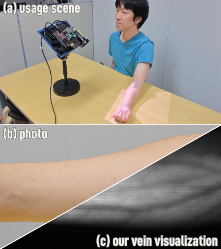
Light provides all our visual information, but it reaches our eyes in different ways. Direct light comes unperturbed, coming straight from the source, whereas indirect light bounces off different surfaces, such as walls or ceilings, before entering our eyes. Extracting information from these two pathways has significant implications in diagnostic imaging and other applications. A multinational collaboration led by Nara Institute of Science and Technology Assistant Professor Hiroyuki Kubo, Arizona State University Assistant Professor Suren Jayasuriya, and Carnegie Mellon University Professor Srinivasa G. Narasimhan has successfully captured and analyzed indirect light with commercially available cameras to create images of blood vessels in living human beings in real time at extraordinary resolution.
While direct light provides information like depth and colour, allowing us to see a person's skin and other superficial features, indirect light, explains Kubo, can reveal otherwise unseen details just under the surface.
"Light on skin has strong subsurface scattering. By capturing indirect light, we can see details of invisible objects underneath the skin like blood vessels," he says.
The new technique exploits the epipolar geometry on which light travels by creating a synchronization delay between the light source and camera. A new nonlinear algorithm developed by the researchers then demultiplexes the information to provide a sharper image.
"Our imaging system illuminates the scene with epipolar planes corresponding to projector rows. We vary two key parameters to capture the light transport: the offset between the projecting row and camera row in the rolling shutter, or synchronization delay, and the exposure of the camera row," explains Kubo.
The result was the imaging of blood vessels at a much finer resolution than standard medical instruments. It also extracted details in circumstances that have been challenging in medical imaging, such as darker toned or hairy skin.
"We captured the basilic and cephalic veins in the inner forearm, which are about 1-5 mm deep," observes Kubo.
The hardware comes at a low cost and is portable. That and the ability to reveal new details of blood vessels non-invasively will improve the quality of care to a wider patient population such as patients who cannot have agents injected into their blood vessels, like children or the elderly.
Overall, Kubo is optimistic that this technique adds to vascular image quality in just about any condition seen in clinics.
"Our method is robust to ambient light, curvatures, and obstructions such as hair darker skin tones," he says.
Programmable non-epipolar indirect light transport: capture and analysis
Hiroyuki Kubo, Suren Jayasuriya, Takafumi Iwaguchi, Takuya Funatomi, Yasuhiro Mukaigawa & Srinivasa G. Narasimhan
IEEE Transactions on Visualization and Computer Graphics (2019)
DOI: 10.1109/TVCG.2019.2946812
Contact information:
Assis. Prof. Hiroyuki Kubo at Division of Information Science, Graduate School of Science and Technology,
Nara Institute of Science and Technology
hkubo@is.naist.jp
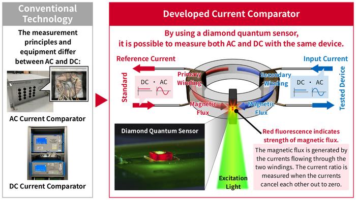
Compact current comparator based on diamond quantum sensing for the unified calibration of AC and DC current measurement instruments Researchers at AIST, in collaboration with the Institute of Science Tokyo and the National Institutes for Quantum Science and Technology, have developed a “current comparator” that uses diamond quantum sensors to accurately evaluate both AC and DC current ratios using the same device setup. A current comparator is a device for the calibration of current...
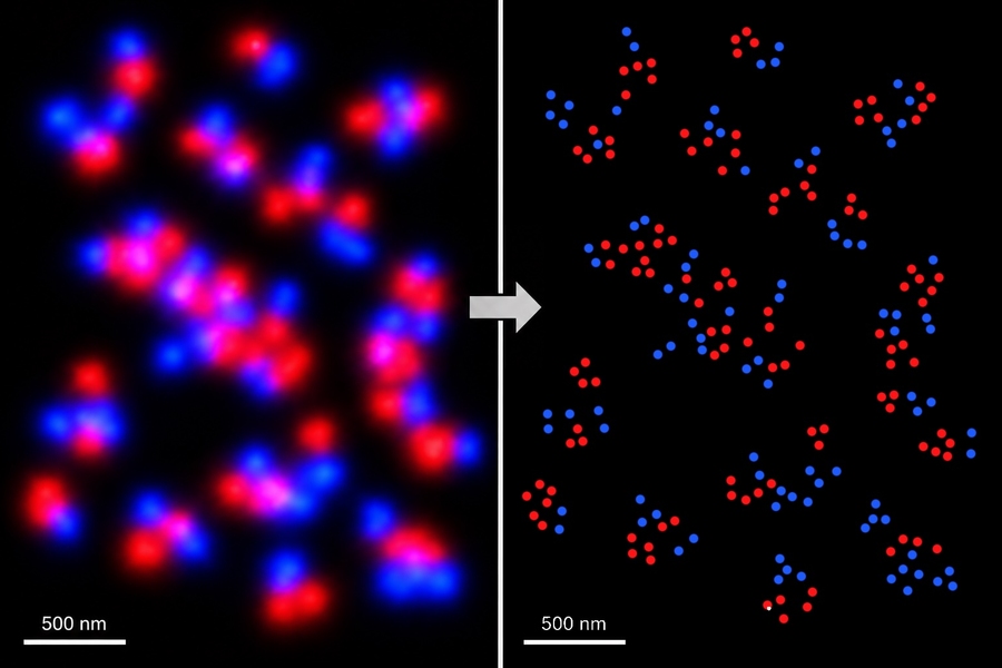
MIT and Broad Institute researchers have developed U-STORM, a super-resolution microscopy platform using engineered upconverting nanoparticles that blink spontaneously and indefinitely. The method achieves 0.6-angstrom localization precision with a single near-infrared laser, enabling multicolor imaging without complex buffers or multiple lasers. This simplifies high-precision molecular imaging for biological applications. Published in Nature Nanotechnology.
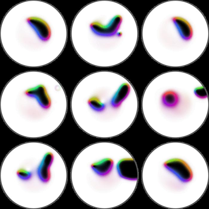
A team of researchers from the Max Born Institute, the Ferdinand Braun Institute, the University of Augsburg, and the Helmholtz-Zentrum Berlin has succeeded in directly imaging the effect of short current pulses on a skyrmion.
