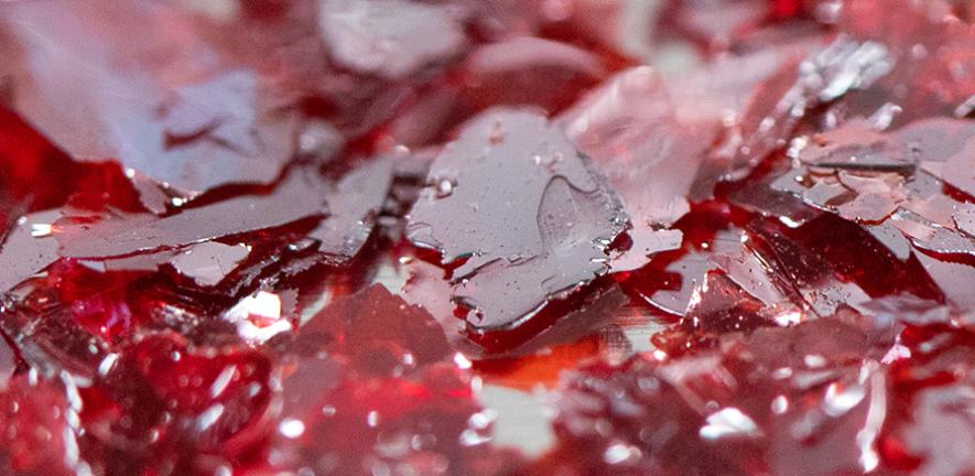
Using X-rays to see inside the human body has revolutionised non-invasive medical diagnostics. However, the dose of X-rays required for imaging is far higher than background levels, due to the poor performance of the detector materials currently available. This can cause harm to patients, and in some cases even cancer.
A team of researchers, jointly led by the Universities of Oxford and Cambridge, have discovered that a solar cell material – bismuth oxyiodide (BiOI) – is capable of detecting X-ray dose rates over 250 times lower than the current best performing detectors used commercially. This has the potential to make medical imaging safer, and open up new opportunities in non-invasive diagnostics, such as X-ray video techniques. Their results are reported in the journal Nature Communications.
“We have developed BiOI single crystals into X-ray detectors that work over 100 times better than the current state-of-the-art for medical imaging,” said Dr Robert Hoye from the University of Oxford, who led the work. “BiOI is nontoxic, stable in air, and can be grown cost-effectively and at scale. We are very excited by the potential BiOI has to make the next generation of non-invasive diagnostics more accessible, safer, and more effective.”
BiOI is a nontoxic semiconductor that absorbs visible light and is stable in air. Owing to these qualities, over the past decade there has been a surge of interest in this material for solar cells (turning sunlight into clean electricity), photoelectrochemical cells (turning sunlight into fuels) and energy harvesting to power smart devices, among many other applications.
BiOI contains two heavy elements – bismuth and iodine – which allows the material to strongly absorb X-rays. However, previous attempts to make BiOI into X-ray detectors were ineffective due to significant energy losses from defects arising from the nanocrystalline nature of the detectors made.
The researchers developed and patented a method to grow high-quality single crystals of BiOI using a scalable vapour-based approach. The low defect density in these crystals led to stable and ultra-low dark currents, which was critical to substantially improve the sensitivity and detection limit of this material to X-rays.
“Showing that these simply-processed, low-temperature grown, stable crystals can give such high sensitivity for X-ray detection is quite remarkable,” said Professor Judith Driscoll from Cambridge’s Department of Materials Science and Metallurgy, who co-led the work. “We began working on this material, BiOI, several years ago, and we find it outshines other rival materials in a range of optoelectronic and sensing applications, when toxicity and performance are considered together.”
The researchers formed an interdisciplinary team to understand why BiOI works so well as an X-ray detector. They used advanced optical techniques to resolve processes taking place over a trillionth of a second, and coupled these with simulations to link these processes with what is happening at the atomic level.
Through this study, the team revealed the unusual way in which electrons couple to vibrations in the lattice. Unlike other bismuth-halide compounds, the electrons in BiOI remain delocalised, meaning that electrons can easily and rapidly move within the lattice of BiOI. At the same time, the unusual electron coupling with lattice vibrations results in an irreversible energy loss channel that would still be present even if the material were defect-free.
The researchers found that these losses can be overcome by cooling down the sample to reduce thermal energy, or by applying an electric field to rip away electrons from the lattice. The latter case is ideally matched with how X-ray detectors operate. By applying a small electric field, electrons can be transported over a millimetre length-scale, allowing the efficient extraction of electrons generated in the single crystals through the absorption of X-rays.
“We have built a microscopic quantum mechanical model of electrons and ions that can fully explain the remarkable optoelectronic properties of BiOI that make it such a good material for X-ray detection,” said Dr Bartomeu Monserrat from Cambridge’s Department of Materials Science and Metallurgy, who co-led the project. “This gives us a roadmap for designing even more materials with similarly advantageous properties.
This work offers important insights into how delocalised charge-carriers can be achieved in bismuth-halide compounds. The researchers are now working on applying these insights to design materials with similarly advantageous properties as BiOI, as well as how to tune the composition of BiOI to improve its transport properties further. They are also working on bringing the unique benefits of BiOI to society by devising routes to increase the size of the BiOI detectors, while preserving the exceptional properties found in single crystals.
Reference
Layered BiOI single crystals capable of detecting low dose rates of X-rays
Robert A. Jagt, Ivona Bravić, Lissa Eyre, Krzysztof Gałkowski, Joanna Borowiec, Kavya Reddy Dudipala, Michał Baranowski, Mateusz Dyksik, Tim W. J. van de Goor, Theo Kreouzis, Ming Xiao, Adrian Bevan, Paulina Płochocka, Samuel D. Stranks, Felix Deschler, Bartomeu Monserrat, Judith L. MacManus-Driscoll & Robert L. Z. Hoye
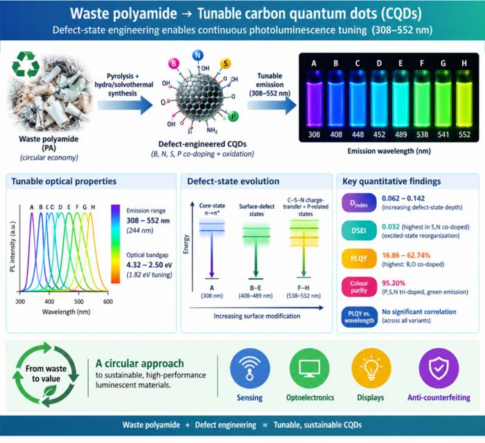
Waste polyamide-derived CQDs show continuous photoluminescence tuning from 308 to 552 nm through sequential defect-state engineering, with optical transition energies decreasing from 4.32 to 2.50 eV. @Christian Ebere Enyoh from Saitama University Carbon quantum dots (CQDs) are fluorescent carbon nanomaterials with potential applications in sensing, optoelectronics, displays, anti-counterfeiting, and environmental technologies. Their optical properties can be adjusted by modifying the carbon...
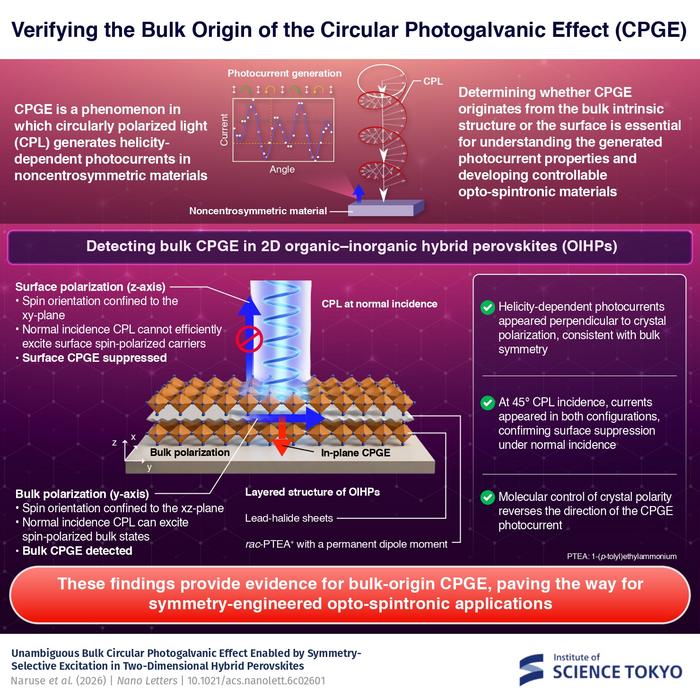
The researchers used circularly polarized light at normal incidence and found that helicity-dependent photocurrents appeared perpendicular to the crystal’s polarization but vanished when measured parallel to it, supporting a bulk origin of CPGE. @Institute of Science Tokyo The circular photogalvanic effect (CPGE), a phenomenon that generates helicity-dependent photocurrents in noncentrosymmetric materials, can originate purely from a crystal's internal structure without contribution from the...
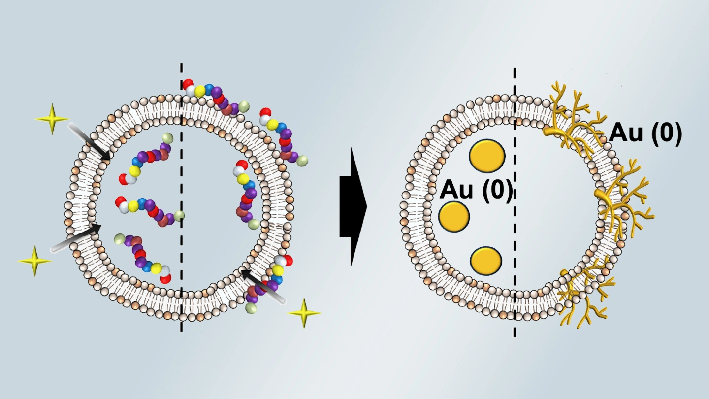
Summary of gold nanoparticle growth programming in liposome based on localization of biomineralization peptide The position of biomineralization peptides within liposomes can influence how gold nanoparticles grow, reports a research team from Institute of Science Tokyo. Peptides localized at the membrane interface promote branched structures, while those confined to the liposome interior favor spherical nanoparticles. The findings offer a new strategy for controlling nanoscale reaction...
