University of Vienna
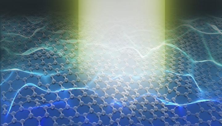
In order to understand advanced materials like graphene nanostructures and optimize them for devices in nano-, opto- and quantum-technology it is crucial to understand how phonons - the vibration of atoms in solids - influence the materials' properties. Researchers from the University of Vienna, the Advanced Institute of Science and Technology in Japan, the company JEOL and La Sapienza University in Rome have developed a method capable to measure all phonons existing in a nanostructured material. This is a breakthrough in the analysis of nanoscale functional materials and devices. With this pilot experiment using graphene nanostructures these researchers have shown the uniqueness of their approach, which will be published in the latest issue of Nature.
Important thermal, mechanical, optoelectronic and transport characteristics of materials are ruled by phonons: the propagating atomic vibrational waves. It is then inferable that the determination of such extended atomic vibrations is crucial for the optimization of nanoelectronic devices. The current available techniques use optical methods as well as inelastic electron-, x-ray- and neutron scattering. Despite its scientific importance in the last decade, none of these methods has been able to determine all phonons of a freestanding monolayer of two dimensional (2D) materials such as graphene and their local variations within a graphene nanoribbon, which are in turn used as active elements in nano- and optoelectronics.
The new limits of nanospectroscopy
An international research team of leading experts in electron spectroscopy led by Thomas Pichler at the University of Vienna, theoretical spectroscopy led by Francesco Mauri at La Sapienza University in Rome and electron microscopy led by Kazu Suenaga at the AIST Tsukuba in Japan, together with the Japanese company JEOL have presented an original method applying it to graphene nanostructures as model: "high resolution electron spectroscopy inside an electron microscope with enough sensitivity to measure even an atomic monolayer". In this way they could for the first time determine all vibrational modes of freestanding graphene as well as the local extension of different vibrational modes in a graphene nanoribbon.
This new method, which they called "large q mapping" opens entirely new possibilities to determine the spatial and momentum extension of phonons in all nanostructured as well as two dimensional advanced materials. These experiments push the limits of nanospectroscopy approaching the limits of Heisenbergs uncertainty principle and demonstrates new possibilities to study local vibration modes at the nanometer scale down to individual monolayers.
New electron nanospectrometer as "table top" synchrotron
"The direct experimental proof of the full spatial and momentum resolved mapping of local vibrations of all materials including even monolayer 2D materials and nanoribbons will enable us to fully disentangle different vibration modes and their momentum transfers at non-perfect structures such as edges or defects, which are extremely important to understand and optimize the local properties of a material", explains one of the leading authors, Ryosuke Senga.
This study of "High q-Mapping Of Vibrations" in the electron microscope opens a new pathway of nanospectroscopy of all materials combining spatial and momentum resolved measurements. This has been the biggest challenge regarding the combination of microscopy and spectroscopy, since the spatial and momentum resolutions are compensated due to the limit of Heisenbergs uncertainty principle. "We believe that our methodology will boost vast research in material science and will push high resolution electron spectroscopy in electron microscopy to the next level, to be envisaged as a true table top synchrotron", says Thomas Pichler from the University of Vienna.
Position and momentum mapping of vibrations in graphene nanostructures
Ryosuke Senga, Kazu Suenaga suenaga-kazu(at)aist.go.jp , Paolo Barone, Shigeyuki Morishita, Francesco Mauri francesco.mauri@uniroma1.it, Thomas Pichler
Nature, 2019
DOI: 10.1038/s41586-019-1477-8
Contact information:
Thomas Pichler
Group Speaker of Electronic Properties of Materials Group at University of Vienna
thomas.pichler@univie.ac.at
Phone: +43-1-4277-51466
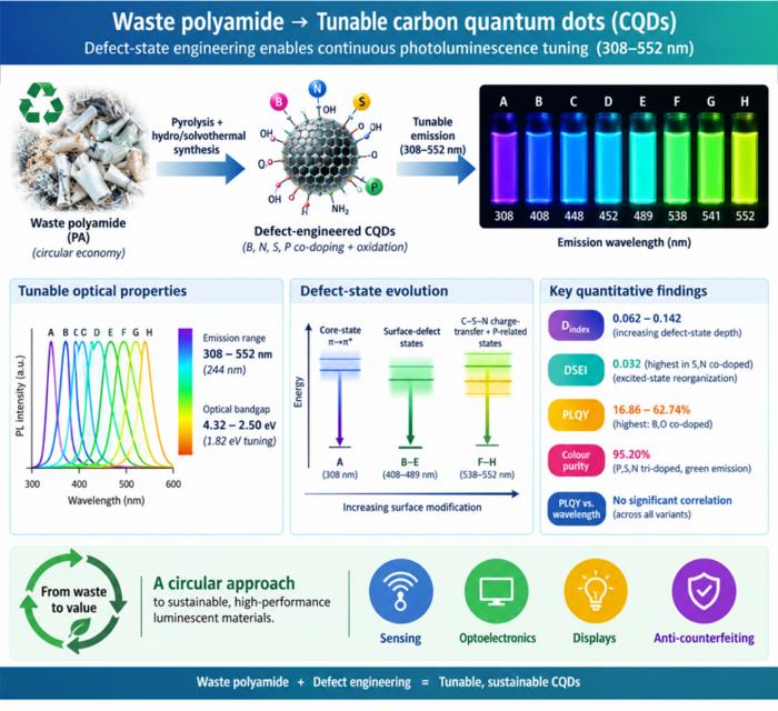
Waste polyamide-derived CQDs show continuous photoluminescence tuning from 308 to 552 nm through sequential defect-state engineering, with optical transition energies decreasing from 4.32 to 2.50 eV. @Christian Ebere Enyoh from Saitama University Carbon quantum dots (CQDs) are fluorescent carbon nanomaterials with potential applications in sensing, optoelectronics, displays, anti-counterfeiting, and environmental technologies. Their optical properties can be adjusted by modifying the carbon...
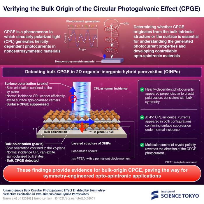
The researchers used circularly polarized light at normal incidence and found that helicity-dependent photocurrents appeared perpendicular to the crystal’s polarization but vanished when measured parallel to it, supporting a bulk origin of CPGE. @Institute of Science Tokyo The circular photogalvanic effect (CPGE), a phenomenon that generates helicity-dependent photocurrents in noncentrosymmetric materials, can originate purely from a crystal's internal structure without contribution from the...
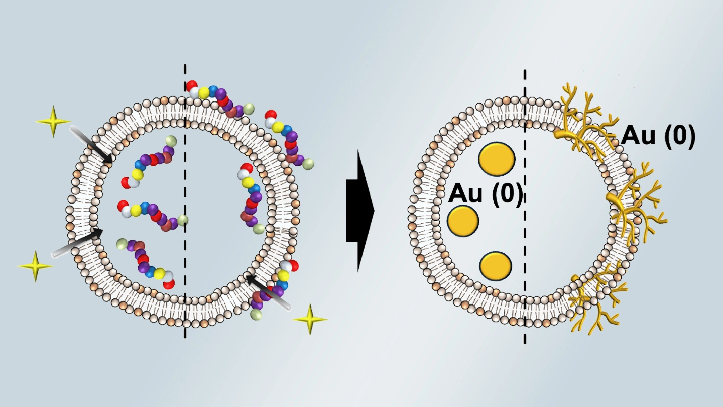
Summary of gold nanoparticle growth programming in liposome based on localization of biomineralization peptide The position of biomineralization peptides within liposomes can influence how gold nanoparticles grow, reports a research team from Institute of Science Tokyo. Peptides localized at the membrane interface promote branched structures, while those confined to the liposome interior favor spherical nanoparticles. The findings offer a new strategy for controlling nanoscale reaction...
